The Decorations of Oregonia gracilis (Decapoda, Brachyura, Majidae)

If you pull up any tire from the edge of the docks at Friday Harbor Laboratories, you’ll find a multitude of organisms — algae, bryozoans, hydroids, tube worms, and tunicates growing on the surface, and many more mobile organisms such as crabs, shrimp, urchins, copepods, and a variety of worms living in the habitat thus created.

DGP_3651

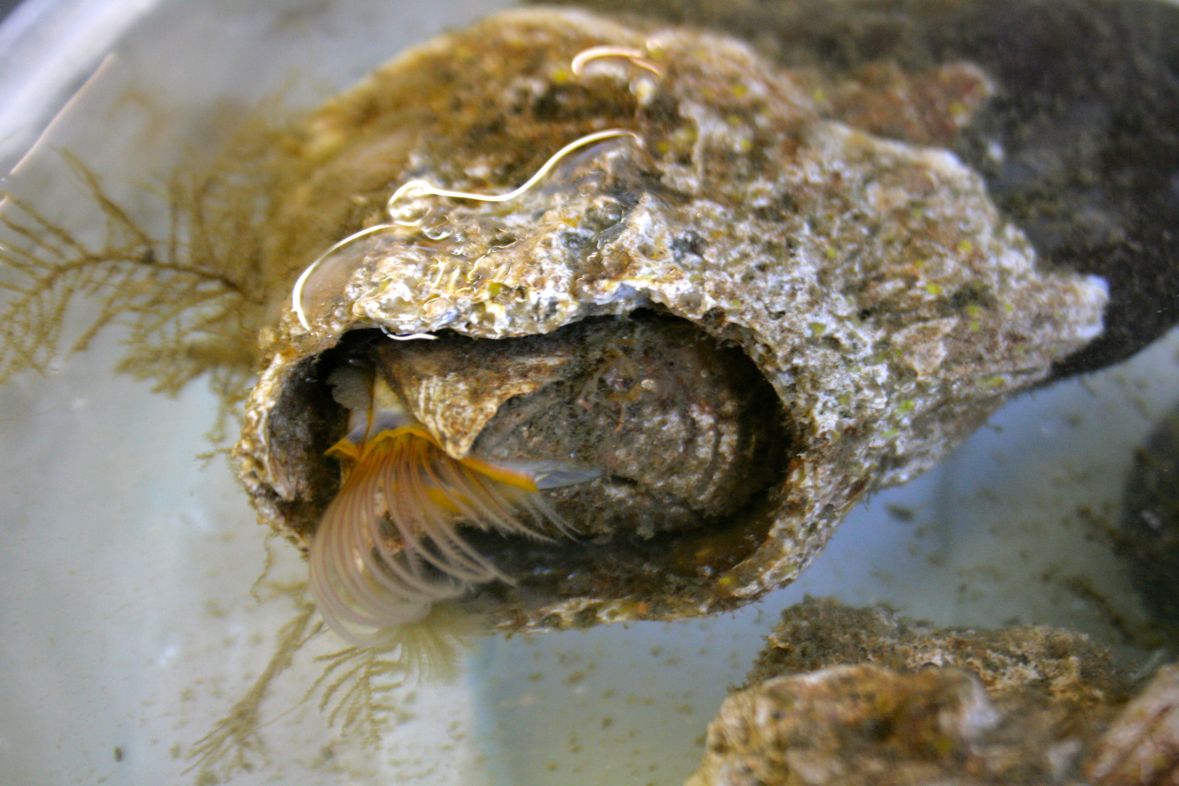
Figure 1 — Mussel (left), limpet (upper right), and rock (lower right), overgrown with a variety of organisms

On the benthos, a similar situation exists — any available substrate, living or not, is swiftly colonized by a variety of organisms. The rock in figure 1, though relatively small, is home to Terebratalia transversa, a brachiopod; red algae; at least four species of encrusting bryozoans; small calcareous tubes which could belong to gastropods or serpulid polychaetes; and two larger worm tubes, one containing its worm (Sabellaria), one containing a hermit crab (Discorsopagurus). Living substrates are likewise colonized; the mussel in figure 1, which was dredged from the same location as the rock, is overgrown by Balanus, the acorn barnacle; T. transversa; and at least two types of thecate hydroids. Finally, the limpet (next to the mussel in figure 1) carries on its back the calcareous bryozoan Heteropora, spionid polychaetes, Balanus, red algae, and an encrusting bryozoan. This sort of overgrowth of some organisms by others is an inevitable consequence of the scarcity of space in the marine benthos. Some, such as cheilostome bryozoans with their grabbing avicularia, combat the encrustation. Some, like the mussels and the limpet, ignore it. Finally, some organisms take advantage of it.

The decorator crab (Oregonia gracilis) is named for the patchwork of organisms which it purposefully attaches onto hooked setae on its carapace. Once emplaced, the organisms establish and grow there. This camouflaging cloak of colonists serves to confuse predators, and the makeup of the decorations is known to vary from one place to another, since these crabs choose their decorations from locally-available materials (Wickstein 1992). Decorator crabs are commonly seen on the support posts of the dock here at Friday Harbor, below the water line, so I went down to the dock with a net and a bucket to catch a few and see what they had attached to their carapaces.

I caught three O. gracilis individuals. My first impression of their decorations was that in contrast to the rock, mussel, and limpet I examined earlier, the crabs’ encrusting biota was much more uniform. While the rock had patches of bryozoans and clumps of worm tubes, and the mussel’s hydroids were concentrated at one end, the crabs had organisms evenly distributed over their carapaces. Furthermore, the community composition of different parts of a crab’s carapace was similar; crabs with bryozoans on their backs also had the same type of bryozoans on their legs (Figs. 2, 4), and a crab with spots of sponge on its back had spots of the same sponge on its legs (Fig. 3). In contrast, the rock and mussel communities were much patchier.

So, what lives on the carapaces of decorator crabs at Friday Harbor? Let’s take a look.

Fig2

Figure 2 — Dorsal view of O. gracilis with Dendrobenia lichenoides, red algae, yellow sponge, Corella willmeriana (transparent tunicate), four hydroids, and diatoms

The first crab I caught (Fig. 2) had attached a large frill of the bryozoan Dendrobaenia lichenoides to its rostrum. Smaller flakes of D. lichenoides were attached to its carapace and legs. Among the bryozoans grew at least four species of colonial hydroids. Diatom chains were stuck among the hydroids in many places; since those were not attached directly to the carapace, I am not sure whether or not the crab did that on purpose. Finally, the crab had finished off its decorations with a few bits of red algae, some unidentified yellow sponge, and a transparent tunicate which it carried along on one leg.

Fig3

Figure 3 — Dorsal view of O. gracilis with Dendrobenia lichenoides, green algae, red algae, and unidentified hydroids

The second crab (Fig. 3), smaller than the first, was thickly adorned with D. lichenoides and a number of hydroids. It added some heterogeneity into its camouflage with some flecks of red and green algae.

Fig4

Figure 4 — Dorsal view of O. gracilis with Dendrobenia lichenoides, yellow sponge, red algae, and green algae

The third crab (Fig. 4), like the first two, had also placed D. lichenoides all over its body. Unlike the first two, however, it had also polka-dotted itself with yellow sponges. It also had a few flecks of red and green algae, but no hydroids that I could find.

While the species composition from one crab’s carapace to another is not identical, a number of similarities emerge. Overall, the crabs appear to have attached leafy or dendritic, neutral-colored, colonial animals over most of their carapaces, and then added smaller patches of brighter-colored materials. The species on their carapaces are all common on local non-living substrates; for instance, it is easy to find Dendrobaenia on the dock tires. Finally, just like the bryozoans, algae, and hydroids on the dock tires and the dredged rocks and mussel, the habitat built by the decorator crabs on their carapaces provides a home for a number of other species; I found a number of amphipods, nematodes, and polychaetes living amongst their bryozoans and hydroids. Thus, while decorator crabs purposefully cultivate sessile organisms for their camouflage, they incidentally attract more mobile organisms as well and thus carry full-fledged communities on their backs.

Nadia Pierrehumbert
University of Chicago

REFERENCES

Wicksten, Mary K. 1993. A review and a model of decorating behavior in spider crabs (Decapoda, Brachyura, Majidae). Crustaceana 64(3).

Diversity of Parapodia in Polychaete Worms

One of the most distinctive features of polychaetes are their parapodia. Parapodia are the paired “legs” of a polychaete that are outgrowths of each body segment. They can have a variety of functions and thus take on a variety of forms. The purpose of this blog post is to explore the diversity in the morphology and function of parapodia in different polychaete families.

The General Parapodium

Parapodia are biramous. They have a dorsal notopodium and a ventral neuropodium. These lobes usually have chaetae (notochaetae or neurochaetae), which are bristles made of chitin and protein. The notopodium can have a dorsal outgrowth called a dorsal cirrus while the neuropodium can have a ventral cirrus. These cirri generally have sensory functions. Parapodia can also have branched outgrowths called branchia which often function as gills.

Errantia

Errantia is one of the two major groups of polychaetes. They generally have large, well-developed parapodia.

Image

Nereididae—Nereis brandti: Parapodia are large and are used for fast crawling and swimming. The dorsal cirri get progressively larger in the posterior direction, with the posterior segments having very prominent lobe-shaped dorsal cirri. that may help with locomotion by increasing the surface area of the parapodia.

Nephtyidae

Nephtyidae—Nephtys: Parapodia are also quite large. Nephtys have rigid, muscular bodies and are active burrowers. Their large parapodia are relatively simple and facilitate movement through substrate.

Glyceridae

Glyceridae—Glycera: Glycerids are generally carnivorous. They have elaborate burrow systems to lie in wait for prey, and presumably use their large parapodia to dig and move quickly. They have a huge eversible pharynx. I observed a red tuft that resembled branchia on the ventral side of the parapodium. However, the literature describes this tuft to be on the dorsal side. Regardless, these filamentous red structures are not true branchiae because they contain no circulatory system, which has been lost in this family. Instead, they are known as coelomic loops, and are red because the coelomic fluid that flows through it contains hemoglobin.

Lumbrineridae

LumbrineridaeLumbrineris: These worms are long, stringy and fairly firm. They have small uniramous parapodia, with only the neuropodium present. Reduction of the notopodium occurs in several families, since the ventral neuropodium is usually more important for locomotion. Like Nephtys, they use their well-developed musculature to burrow. They are generally found in masses under rocks.

Polynoidae

PolynoidaeHalosydna brevisetosa (the scale worm): This polychaete family has scales, called elytra, that are secreted from parts of the notopodium. This particular species have 18 pairs of elytra. Parapodia are large, with a wide bunch of chaetae on the neuropodium. The notopodium is also reduced and the dorsal cirri are present on every other parapodium. They are found sticking to the undersides of rocks.

Eunicidae

EunicidaeEunice: These worms construct tubes in the sediment, from which they extend to feed. They have long branchiae for respiration. The notopodium is reduced to just the dorsal cirrus.

Onuphidae

Onuphidae—Diopatra: These worms live in tubes that can get very deep and deoxygenated. Ours was found in a tube that was arm’s-length deep into the mud. Thus, they have very large gills anteriorly for respiration but the gills get progressively smaller down the length of the body.

Sedentaria

Sedentaria is the other major group of polychaetes. They generally have smaller, simpler parapodia than Errantia. Two major groups of these sedentary worms are Canalipalpata and Scolecida.

Canalipalpata generally live in tubes.

Terebellidae

TerebellidaeThelepus (the spaghetti worm): Terebellids live in tubes and stick their head tentacles out to feed. The neuropodium is a compressed, long, thick structure that extends ventrally. When the neuropodium takes this form, it is called a torus. The tori bear hook-like chaetae called uncini, while the notopodium is smaller and bears the regular, long chaetae. The uncini help anchor the worm in its tube, while the long chaetae help it to move around in the tube.

Sabellidae

Sabellidae—Eudistylia (the feather duster worm): The thoracic and abdominal sections of this worm are distinct because of chaetal inversion. In the thorax, the notopodium has the long chaetae while in the abdomen, the neuropodium has it. The thoracic neuropodium and abdominal notopodium are tori and have uncini.

Cirratulidae

Cirratulidae—Cirratulus: The most characteristic feature of this worm are the long branchia, which function in respiration.  Cirratulids live in mud or sand underneath rocks. Though they don’t have very active lifestyles, they still seem to need lots of gills. I observed they tend to tangle themselves up in their gills. The notopodium and neuropodium are reduced to just the chaetae.

Scolecida are earthworm-like polychaetes.

Arenicolidae

Arenicolidae—Abarenicola pacifica (the lugworm): These worms are deposit feeders. Because they live in burrows, oxygen can be limited so their parapodia have large branchia. Like the Lumbrineris and Nephtys, they rely on musculature to burrow. Their neuropodia are tori.

Andrea Wong

University of Washington

Epidermal Chromatophore Activity in Octopus rubescens (Mollusca, Cephalopoda)

Of the chromatophore variations throughout the animal kingdom, those within the class Cephalopoda are unique in that they are not controlled via the endocrine system, but rather exclusively by direct innervation. These chromatophores include pigment granules contained within a sac, called a cytoelastic sacculus, surrounded by radial muscles. When contracted these radial muscles expand the sacculus, which provides the epidermis with color characteristic of the pigment. Being covered by these chromatophores, cephalopods are capable of remarkable feats of camouflage.

Image

Fig. 1. Diagram showing the anatomy of a Loligo opalescens chromatophore organ (Cloney and Florey, 1968).

The particular species in discussion here is Octopus rubescens, a local species of the Pacific Northwest. The O. rubescens was unintentionally retrieved while collecting a dead bivalve shell on a dive in the San Juan Islands. The specimen had made its lair out of the shell, and went unnoticed until it was brought back to the lab. The specimen died shortly thereafter.

In the process of dissecting the mantle of the specimen, respiratory activity was still evident. Ethanol was added to ensure death. While observing the epidermis under a dissecting scope, the reduced chromatophores began to show activity.

Image

Fig. 2. Posterior side of rubescens during dissection of the mantle, prior to chromatophore activity.

When the radial muscles surrounding chromatophores are relaxed, the elasticity of the tissue retracts the sacculus (Messenger, 2001).  The deceased specimen appeared mostly white to the naked eye (see Fig. 2). It was unexpected to find that individual chromatophores, when observing the epidermis at this magnification, were expanding—the radial muscles around them were contracting. As the solution gradually increased in temperature, this activity increased in frequency and magnitude. Eventually, the posterior side of the specimen was ablaze with changing colors visible to the naked eye.

Video 1. 19:26, solution temperature 12.5°C                               

Video 2. Magnified. 19:32, solution temperature 12.5°C.

Pigment within the sacculus generally falls toward higher wavelengths of the visible spectrum. Yellow, orange, red, and brown characterized the range found on the O. rubescens (see Fig. 3). These pigments are typically derivatives of tryptophan (Messenger 2001). The activity was visible in waves, and seemed to involve one chromatophore color at a time. Considerable overlap between individual chromatophores was observed when contracted, which, when seen from a distance, could allow further ranges of color. This, along with light reflecting iridophores and the ability of the octopus to contort its epidermis to varying textures, probably aids the octopus in camouflage.

Image

Fig. 3.   Magnified epidermis of O. rubescens as chromatophore activity escalated.

The specimen was cooled via refrigeration and addition of seawater to the solution. This was done to minimize time between observations so as to minimize natural degeneration as a factor for reduced activity. At a solution temperature of 7.5 °C, 36 minutes after start of cooling, the specimen was again observed. The chromatophore activity had perceptibly reduced. This observation supports that this chromatophore activity increases with higher temperatures.

 Video 3. 20:13, solution temperature 7.5°C.

This activity was known by physiologists as “Wandernwolken” (“wandering clouds”) in the nineteenth century. Messenger explains this phenomenon as correlated with the death of the innervating cells while the surrounding muscles endure, and waves of color as was observed here could be induced via anaesthetization with ethanol. These waves tend to propagate through “classes” of chromatophores, such as similar color or age. This activity lends support to a form of network link between chromatophores. (Messenger, 2001).

Dominic Sivitilli

University of Washington

References:

Cloney, R. A. & Florey, E. (1968). Ultrastructure of cephalopod chromatophore organs. Zeitschrift für Zellforschung 89, 250-280.

Messenger, J. B. (2001), Cephalopod chromatophores: neurobiology and natural history. Biological Reviews, 76: 473–528. doi: 10.1017/S1464793101005772

Cell circulation in branchiae of Glycera americana

Perhaps to many it may come as a surprise that the glycerid polychaete is a fascinating subject to study. What might seem like a simple worm at first glance, it comes in at about 7 inches in length with a silver sheen on the somewhat pinkish body, and flaunts a gnarly proboscis containing four black jaws used in feeding.

Glycera pic
Fig.1 Glyceridae americana

Expecting the generic annelid characteristics with the particular worm I was looking at, I was very quickly drawn to the parapodia and branchia along the sides of the animal. The parapodia are lateral outgrowths along the worm’s body that are involved in respiration and locomotion while the branchiae are dorsal extensions of the parapodia, essentially the gills.glycera body:parapodia

Fig.2 Illustration of G. americana with a close up of a parapodium and its associated branchia

The branchiae clearly had movement of cells occurring inside of them when viewed under the dissecting microscope, and I was curious to see what kind of cells were circulating and how they were being driven. I snipped off one of the branchiae from an anterior segment of the worm and prepared a slide to view it under the compound microscope. The action going on inside just the single outgrowth was really exciting; numerous, small, red disc-shaped cells, fewer large white disc-shaped cells, and several small circular green cells were circulating around in what seemed to be a patterned flow.brachia pic

Fig.3 Even closer view of the branchia with the direction of cell flow of the three different cell types observed
Although the branchia was removed from the body and therefore possibly altered the movement of blood, the cells continuously flowed up one side and down the other of each individual groove or inlet of the branchia, with the smaller red cells passing around the larger white cells. The red cells seemed to move the fastest, with the smallest extracellular green cells a bit slower, and the large disc-shaped white cells barely moving by. The flow itself was not pulsating, and as polychaetes do not have a heart or cardiovascular system, ciliary movement was the force smoothly pushing the cells along the coelomic cavity. Also amazing was the fact that the movement of cells continued after the branchia was removed from the body for about 40 minutes. The video below shows the circulation of cells.

The next thing I was interested in finding out was the likely function(s) of each cell type, assuming at least one type was a part of the blood system. In their book Polychaetes, Greg Rouse and Fredrik Pleijel confirm that “Glyceridae lack a circulation system, the respiratory pigment is located in disc-shaped erythrocytes floating in the coelom” determining that one of the cells are indeed involved in respiration and that cilia are the driving force rather than muscular pulsing (112). An email from Jenna Moore, a polychaete expert, explained that the “glycerids [do] have hemoglobin…[and the] green [cells] could be chlorocruorin, another blood pigment” however the large translucent white cells still remain to be identified. The glycerids display a great visual of blood flow in the worms through their branchia and seeing all of this and how it worked under the microscope was great. Further research in this area of blood cells and the circulatory pathways involved in invertebrates would be fascinating.

Ashley Shiner

University of Washington

Literature Cited:

Pleijel, Fredrik and Greg Rouse. Polychaetes. New York: Oxford University Press, 2001. 112. Print

Sampling the Diversity of Alpheid Shrimp Microhabitats: Examples from the Pacific Northwest and the Indo-West Pacific

Alpheidae is a family of caridean shrimp that contains the snapping shrimp genera Alpheus and Synalpheus, in addition to a few other non-snapping genera. Although the most diverse genus in this family (Alpheus) typically inhabits nearly all tropical and sub-tropical shallow water marine habitats, alpheids in general can be found in many diverse places, from mud flats to coral reefs to seagrass beds, including in temperate waters, with some individuals even inhabiting deep-water Caribbean locales.

I experienced a small portion of this habitat diversity during two field seasons studying Alpheus genetic and cryptic diversity in Bali and Sumatra, Indonesia. Most of our samples were collected from the interstices of dead Pocillopora spp. coral heads (Fig. 1) that were excavated from the reefs, and all others were taken from the surfaces of our Artificial Reef Monitoring Structures (ARMS) (Fig. 2) that were placed on the reefs the year before. During this time, we collected shrimp from 47 unique evolutionarily significant units (ESUs) from many different types of microhabitats on both the ARMS and the coral heads.

Figure 1: Dead Pocillopora coral head taken from a reef off the shore of northern Bali, Indonesia. All Alpheus samples taken from the head were found in the interstices of the coral (from both living and dead colonies), inside of algal tubes hanging from the head, or within caverns excavated inside of the skeleton itself.

Figure 1: Dead Pocillopora coral head taken from a reef off the shore of northern Bali, Indonesia. All Alpheus samples taken from the head were found in the interstices of the coral (from both living and dead colonies), inside of algal tubes hanging from the head, or within caverns excavated inside of the skeleton itself.

Figure 2: Artificial Reef Monitoring Structure (ARMS) being deployed on a reef flat in Bali, Indonesia. All Alpheus samples collected from the ARMS were pulled from the plastic green scrubby shown here as the uppermost tier of the structure.

Figure 2: Artificial Reef Monitoring Structure (ARMS) being deployed on a reef flat in Bali, Indonesia. All Alpheus samples collected from the ARMS were pulled from the plastic green scrubby shown here as the uppermost tier of the structure.

One of the many sampled microhabitats was within the crevices of living Pocillopora spp. corals; A. lottini is a mutualistic symbiont of corals of this genus, trading its defensive capabilities in return for shelter (Fig. 3). It has also adopted cryptic coloration patterns for additional protection from predators.

Figure 3: A. lottini individual collected from a section of living Pocillopora coral growing over a dead coral head. It is a mutualistic symbiont of the living corals and its coloration is cryptic among the colony's interstices.

Figure 3: A. lottini individual collected from a section of living Pocillopora coral growing over a dead coral head. It is a mutualistic symbiont of the living corals and its coloration is cryptic among the colony’s interstices.

Another microhabitat we sampled was inhabited by A. frontalis: shrimp of this species formed tubes from algal mats that grew over the dead corals (Fig. 4).

Figure 4: A. frontalis individual collected from within algal tubes it made from algal mats overgrowing the dead coral skeleton.

Figure 4: A. frontalis individual collected from within algal tubes it made from algal mats overgrowing the dead coral skeleton.

A. obesomanus, which is recognizable by the distinctive hammer-like dactylus on its major claw, was found within tunnels it had excavated inside the coral’s limestone skeleton using this modified dactylus (Fig. 5).

Figure 5: A. obesomanus ovigerous female collected from a cavity she excavated inside of the dead coral skeleton using the hammer-like dactylus of her major cheliped. She is shown with a bopyrid parasite inside of her carapace.

Figure 5: A. obesomanus ovigerous female collected from a cavity she excavated inside of the dead coral skeleton using the hammer-like dactylus of her major cheliped. She is shown with a bopyrid parasite inside of her carapace.

We also observed shrimps in the genus Synalpheus living with sea lilies – individuals we collected from crinoids of different colors were found to be of the same species complex (S. stimpsonii), but were cryptically colored to match their hosts (Fig. 6).

Figure 6a: Ovigerous female of the S. stimpsonii complex collected from the oral surface of the central disk of a yellow crinoid.

Figure 6a: Ovigerous female of the S. stimpsonii complex collected from the oral surface of the central disk of a yellow crinoid.

Figure 6b: S. stimpsonii male collected alongside (6a) on a yellow crinoid.

Figure 6b: S. stimpsonii male collected alongside (6a) on a yellow crinoid.

Figure 6c: Ovigerous female of the S. stimpsonii complex collected from a black crinoid on the reef flat.

Figure 6c: Ovigerous female of the S. stimpsonii complex collected from a black crinoid on the reef flat.

During my time on San Juan Island, I was also fortunate enough to collect a single alpheid representative from the mud flats of Argyle Bay during low tide on July 10, 2013. This individual, of the species Betaeus harrimani, was a commensal of the mud shrimp, Upogebia spp., and was collected from the caverns excavated by the much larger shrimp. The individual I encountered was an ovigerous female that was 4.9cm long (Fig. 7).

Figure 7: Ovigerous B. harrimani female collected from the cavern system dug by Upogebia in the mud flats of Argyle Bay on San Juan Island.

Figure 7: Ovigerous B. harrimani female collected from the cavern system dug by Upogebia in the mud flats of Argyle Bay on San Juan Island.

Betaeus is a minor genus of Alpheidae, containing around 28 species, and in the Pacific Northwest, there are only representatives from two of these species. What surprised me most was that this genus does not possess asymmetrical chelipeds and cannot snap. During my time working with the Betaeus female, I found that instead of snapping to defend herself, like all of the other alpheid shrimp I’ve worked with, she held out her symmetrical pincers in a defensive stance that resembled that employed by many Brachyuran crabs when threatened.

While observing her behavior, I also noticed that she used both her pleopods and her pereopods to locomote. This struck me as odd because I knew she was ovigerous and that caridean shrimp attach their eggs to their pleopods via an adhesive substance. Upon a closer look at her swimming legs, it appeared that the eggs were attached instead to her sternites since the legs and the eggs moved independently while she swam (Fig. 8). In terms of both aerating the eggs and having them remain attached to the body of the mother during development, it would seem as if having the eggs attached to the sternites instead of the swimming legs would be evolutionarily more advantageous.

Figure 8: Left lateral of B. harrimani female. While the pleopods are stationary, the eggs appear to be attached to the legs themselves, but while swimming, the eggs move independently of the pleopods.

Figure 8: Left lateral of B. harrimani female. While the pleopods are stationary, the eggs appear to be attached to the legs themselves, but while swimming, the eggs move independently of the pleopods.

To conclude, in terms of habitat similarities between all of the alpheids I’ve collected, I would hypothesize that shrimp in this family prefer to live interstitially; every shrimp I have collected was found in some sort of nook, be it made of coral, algae, sponge tissue, echinoderm appendages, plastic (in the case of the “scrubby” layer on the ARMS), or gravel. When removed from this habitat and placed into a dish lacking any sort of overhead coverage, each shrimp tended to display thigmotaxis towards the edges of the container. So although the diversity of alpheid habitat preference is great, environmental coverage appears to be the thread that ties them all together.

Victoria Morgan; Cornell University c/o 2013

Color variation in Hemigrapsus oregonensis (Arthropoda, Pancrustacea, Malacostraca, Brachyura)

crab anatomy

Fig. 1: Anterior-dorsal view of Hemigrapsus oregonensis with labeled external anatomy

Though Hemigrapsus oregonensis is commonly known as the Green Shore Crab, it does not always seem to be particularly green. This small crab (fig. 1), found along the Pacific coast from Alaska to southern California, can be any number of colors (see fig. 2). Among 67 specimens collected from Argyle Creek on San Juan Island, WA, and analyzed at FHL, the following color patterns emerged: the crabs seemed to have a solid color on their carapace with a distinct pattern of spots or shapes in another color over this. The solid colors in the sampled individuals were white, red, yellow, grey-blue and many shades of green. Often the carapace color would be a blend or mixture of two or three of these colors. The patterns were usually in black or green, often with small white spots. The setose pereopods of the crabs, which distinguishes them from the similar looking Hemigrapsus nudus, were often green with black and white spots, though they could be any color. Chelae were often white.

Image

Fig. 2: Photos of selected crabs’ carapaces. All collected crabs fit into one of these 28 categories. Of 67 specimens, 28 color morphs were found which suggests that there are likely many more variations out there.

The color variation among this species could be due to genetic variation, sexual dimorphism, environmental factors during or after development, or could change with age or molting stage of the crab. This last hypothesis is supported by evidence that was fortuitously gathered when one crab molted while it was being photographed. The crab was originally beige with maroon and white spots, but after ecdysis the individual emerged with a bright green carapace and the exact same spot pattern as before. This could indicate that color changes with new molts, or that as the crab approaches its molting period, the carapace fades. However, many individuals were lighter than the crab that molted, and did not molt after seven days in the lab (though the environment could have prevented them). Some molted carapaces were found of various colors, many bright red, which seems to contradict the hypothesis that carapace brightness and color are indicative of molting stage.

Image

Fig. 3: Crab molting. This process took about one minute.

Color also does not seem to depend on age or sex. Individuals were collected from 0.8-2.3 cm, presumably of different ages, with no evidence of a progression or trend in carapace color. There also seemed to be no difference in the colors or patterns between males, females or gravid females. As for habitat, the crabs live among rocks that are just as diversely colored as they are, which likely supports carapace color variation. Crabs did not seem to live under rocks that resembled themselves, and differently colored crabs were often found living under the same rock. Environmental factors likely influence color, but apart from habitat, it is hard to know many details of each crab’s environment, such as their developmental circumstances and diet. Genetics must have some effect on the color, and likely determines the pattern on the carapace because the molting crab kept the same pattern even when its color changed after ecdysis. Most likely, some combination of the above factors must influence carapace color and pattern of Hemigrapsus oregonensis crabs; more research is required to unravel this mystery.

Image

Fig. 4: Habitat of Hemigrapsus oregonensis at Argyle Creek on San Juan Island

Rachel Folz- University of Chicago

Naked and exposed spaghetti worms, Eupolymnia heterobranchia (Annelida, Canalipalpata, Terebellida, Terebellidae)

The first worm that comes to peoples’ minds is often the earthworm. Unknown to many non-biologist and marine scientists (and non-worm enthusiasts), the oceans are teeming with a diversity of worms and some of these worms are the most curious and beautifully colored inhabitants of the marine world.

A spectacular group of these animals are the spaghetti worms of the family Terebellidae. True to their name, these worms appear to be a splay of spaghetti noodles on the seafloor. Terebellids live in tubes or burrows on the seafloor and spread their spaghetti-like tentacles (palps) across the sediment surface from the opening of their tube. A terebellid of approximately 6 cm can have dozens of tentacles over 15 cm long (Figure 1). Each tentacle has a longitudinal central groove lined with cilia. The tentacles feel the seafloor for materials that will help construct a sturdy tube or burrow, such as shell fragments, small rocks, and mud that stick to their body with a secreted mucus. Tentacles also search surface sediment for tasty organic matter. The worms bring particles and food back to the burrow via the cilia in a manner similar to a conveyor belt.

Image

Figure 1. Terebellid with tentacles extending 360° about its body. Dashed ellipse outlines the extent of the tentacles’ reach. The tentacles spread covers approximately 518 cm2, estimating the radii of the ellipse is 15 cm and 11 cm. The body length of the terebellid is 6 cm, not including the tentacles or fire red branching gills.

Because the tentacles serve these two purposes, I tested the hypothesis that terebellids will preferentially extend tentacles and move their body toward sediment rich in nutrients or materials useful in the construction of their tubes. To test this hypothesis, I placed two individuals of the species Eupolymnia heterobranchia, collected from Argyle Lagoon, San Juan Island, WA, in a sea table cleaned of debris, sediment, and other organisms (with the exception of a persistent sea anemone). At one end of the table, I lined the walls with a boarder of sediment, approximately 7 cm wide. The worms were removed from their self-constructed mud tubes and placed naked in the sea table. Each spaghetti worm was positioned to face their own sediment wall at a distance of 10 cm (Figure 2).

I observed the worms every 15 minutes for an hour and both of the worms’ movements about the sea table were random. Neither worm transported sediment along their tentacles for feeding or to construct a new body tube during the period of observation. Moreover, the tentacles did not extend in a preferential direction toward sediment; instead tentacles surrounded the body in 360° (Figure 1). Potential reasons for the lack of interest in the sediment include: sediment did not suit the habitat type of the worms, animals were in shock after being stripped of their outer tubes, and/or no predators were present in the sea table to encourage the worms to build tubes. Although I observed no particle transport along the tentacles, these two terebellids did show an unexpected amount of movement (~2 cm/min) about the sea table for being tube/burrow dwellers (Figure 2). However, it remains unclear if this movement was via cilia or body contractions.

Image

Figure 2. Photos of the two terebellids (A & B) in the sea table experimental set-up at their initial placement and for each sequential 15 minutes for the following hour: t0, t15, t30, t45, and t60 (top to bottom). The dimensions of the sea table are 60 cm wide by 120 cm long. The blue and white rulers are both 15 cm for scale. Note the distance covered by these worms in the span of 1 hour: estimated average velocity for these two terebellids is 1.9 cm/min, ranging between 0.5 – 6.0 cm/min.

Caitlin Keating-Bitonti, Stanford University

Feeding Behavior of Balanus nubilus

Balanus nubilus (Arthropoda, Crustacea, Thecostraca, Cirripedia, Thoracica, Sessilia)

The giant barnacle, Balanus nubilus, is a sessile organism that attaches itself on a chosen substrate and remains there for the duration of the organism’s lifespan (Figure 1). They do not leave their shell, yet open it up for life functions such as: feeding, spawning, and respiration. They are suspension feeding animals that use their feet to extract food from the water column. My observations are based upon organisms with apertures or openings that are around 4cm in length. The giant barnacle has long and short cirripedia showing a clear distinction between the two (Figure 2). Under the short cirri are feeding appendages composed of maxillae and a mandible, which looks very sturdy in comparison to the cirri (Figure 3).

Figure 1. Balanus nubilus with open scutum opercular plates.

Figure 1. Balanus nubilus with open scutum opercular plates.

Figure 2. View of cirripedia, feeding appendages of Balanus nublius.

Figure 2. View of cirripedia, feeding appendages of Balanus nublius.

Figure 3. Dissected Balanus nubilus' feeding appendages, a view into the mouth.

Figure 3. Dissected Balanus nubilus‘ feeding appendages, a view into the mouth.

When feeding, the giant barnacle first opens its operculum and then extends and uncurls its cirripedia into the water column (Figure 4). The cirri still retain a slight curve when fully extended into the water column. I found when orienting the water flow from the sea table in the test barnacles direction, verses turing the water flow off, the feeding rate changed. The giant barnacle extends its cirri more frequently when there is less water movement. This appears to increase the water flow around the cirri and helps the barnacle to feed more efficiently. When there is ample water flow, barnacles seem to keep their cirri extended into the water column for longer periods, therefore beating them less frequently. The cirripedia can be swiveled to either side once extended (Figure 5). This action seems to better orient the cirri and face into the current’s flow. Smaller barnacles species feed more rapidly than larger barnacle species. They frequently extend their cirri at a rate of more than once per second whereas larger barnacles beat their cirri at a slower rate.

Figure 4. Ventral view of Balanus nubilus feeding with cirripedia.

Figure 4. Ventral view of Balanus nubilus feeding with cirripedia.

Figure 5. Ventral view of Balanus nubilus orienting cirri in the water column

Figure 5. Ventral view of Balanus nubilus orienting cirri in the water column

Barnacles are likely to stop feeding and narrow the operculum if they sense a predator above them. They seem to most likely sense predators by an interruption of light and shadows being cast over them. An agitated barnacle is timid to readily feed again and may cautiously take many minutes to resume feeding activity.

If a barnacle is poked along the soft tissue under the edge of the operculum, it swivels its operculum around the aperture in order to hide the soft and vulnerable tissue from a predator (Figure 6). On large barnacles this sometimes means they expose a vulnerable area of soft tissue on the other side of their opercula. The swiveling movement seems very quick for such a large barnacle.

Figure 6. The soft tissue of Balanus nubilus exposed.

Figure 6. The soft tissue of Balanus nubilus exposed.

Bailey Navratil
University of Washington

Introvert Behavior in Phascolosoma agassizii (Sipuncula)

Figure 1: Phascolosoma agassizii with its introvert fully evaginated from the body cavity (coelom). At the most anterior end of Phascolosoma spp., a notch in the crown of red tentacles indicates the dorsal orientation (Kozloff, 1990).

Figure 1: Phascolosoma agassizii with its introvert fully evaginated from the body cavity (coelom). At the most anterior end of Phascolosoma spp., a notch in the crown of red tentacles indicates the dorsal orientation (Kozloff, 1990).

One of the major features of the phylum Sipuncula, or the Peanut Worms, is the presence of an introvert extending from a round, vermiform trunk.  The introvert has two pairs of retractor muscles that connect the apical portion of the introvert (which bears the tentacles) to the inner trunk body wall. This allows the animal to retreat into the coelomic cavity or extend its introvert and nearly double its length. To extend its introvert, the retractor muscles relax; this allows the internal pressure within the coelom to force the introvert outwards. (Kozloff, 1990)

Phascolosoma Internal Anatomy

Figure 2: Diagram shows a pictorial representation of the internal anatomy of P. agassizii. Two pairs of large retractor muscles attach low within the trunk body wall and allow for retraction of the introvert. The dissection this image reflects is the result of cutting along the dorsal body wall.

The specimen I observed was collected July 9th, 2013 at Argyle Beach on San Juan Island, WA. My specimen was found under a rock in a cobble slew connecting the Argyle Bay to the Argyle Lagoon. I observed this specimen in both its habitat and in a water table. My observations focused on the morphology and behavior of the introvert.

I have found there are three major methods in distinguishing trunk from introvert. The first and most obvious method is to observe the extent of invagination by the introvert. The second method in distinguishing the two features is by an abrupt narrowing of the body from trunk to body. Unfortunately, when the animal is relaxed, this difference in body width can be difficult observe. In the specimen I observed, Phasolosoma agassizii (Kozloff, 1974), the introvert could be distinguished from the trunk by the stripe pattern emerging and becoming prevalent as you moved anteriorly. A striping pattern was readily visible on the introvert, whereas the trunk was more or less spotted.

The behavior of the introvert is variable depending on the environment. When the animal is placed on a flat surface in a saltwater tank, it will evert and withdraw its introvert frequently in what superficially looks like a blind animal’s “sniffing” behavior. If given enough time, P. agassizii will move, using a series of rolls and tumbles assisted by the introvert, to a rock or sediment. It will then use its introvert to anchor itself underneath the rock, or in the sediment, and bury itself (anterior positioned down in the case of sediment burrowing). Often, I would find my specimen hiding underneath a rock. Similarly, P. agassizii in the field can be found under rocks anchored to the underlying rocks and sediments.

Side-by-side with scale copy

Figure 3: Image shows the discrepancy in length of the same specimen of P. agassizii when its introvert is near fully invaginated (left, 6 cm) and fully evaginated (right, 11 cm).

When the animal is underneath an object, it will remain there with no obvious effort to escape. Rather, it will evert its introvert completely. Moreover, despite it appearing to be in a rather uncomfortable situation, if one were to remove P. agassizii from its “pinned” state, it will ultimately roll, tumble, and pull its way back and resume being pinned with its introvert fully extended. Not only do these behaviors show the versatility of the introvert, they are also indicative of the durability of the body wall as the animal positions itself tightly under large stones.

Introverts are expressed in other animal phyla, so it appears to be a rather efficient organ. Morphologically, the introvert allows the animal to keep its trunk, which is full of essential viscera, stationary while the introvert may be extended and retracted as it searches for food in sediments. It also allows for rather unique forms of locomotion and a method to hide a large portion of its body from disturbance. Despite the basic morphology of the introvert, the behaviors are diverse and contribute to a very unique organism.

Michael Tassia

University of Washington

Citations:

Kozloff, Eugene N. “Phylum Sipuncula: Peanut Worms.” Invertebrates. Philadelphia: Saunders College Pub., 1990. 345-347. Print.

Kozloff, Eugene N., Linda H. Price, and Eugene N. Kozloff. “Phylum Sipuncula.” Marine Invertebrates of the Pacific Northwest. Seattle: University of Washington, 1987. 181. Print.

The Dissection of Abarenicola claparedi (Annelida, Polychaeta)

On a quiet sunny day, my class went to Eagle Cove, San Juan Island to dig Abarenicola claparedi for our first dissection. The best and easiest way to find these worms is looking for their burrow openings and fecal castings on the surface of the sand. Then we used the shovels to dig as much sand as we could and searching them.

lugworm2-2010

Figure 1. Photo of A. claperedi. Anterior right and posterior left.

1

Figure 2. Drawing of live A. claperedi. Dorsal side. Anteiror left, posteiror right.

Abarenicola claparedi belongs to the class Polychaeta, phylum Annelida. Figure 1 and 2 shows the appearance of the A. claperedi. The whole worm is 20cm long, and1cm wide at the head. The first segments, including the head, are green, followed by yellow body segments. The color of the mouth is red and the anus is orange-red. There are red branchia on both sides of the middle region.  A. claperedi lives in borrows in the sand. It swallows the sediment and feeds on the organic material. Its feces can be seen as sandy ropes outside of the opening to its burrow. Meanwhile, the sand will collapse a little because of the swallow.

2

Figure 3. Drawing of dorsal view with branchia. At 15x magnification of dissecting scope. Also shows the complete part of parapodium

Looking closely to the skin, there are orange dots covering the skin. The parapodium consists of a ventral neuropodium with chaetae and a dorsal brachium (gill).  Figure 3 shows the details of the branchia. Lots of branches increase the surface area for gas exchange. The red color is respiratory pigment in capillaries that go through each branch, transferring oxygen while “breathing”. Observed at 15x magnification on the dissecting scope, the skin of the A. claperedi is translucent yellow: you can see the gut but not clearly underneath the skin.

3

Figure 4. Drawing of a maturing spermatocyte finding in the body liquid of A. claperedi. At 400x maginification of compound scope.

Finishing observations of external features, we started to dissect the A. claperedi. We cut a small hole with fine scissors and sucked some liquid flowing out from the worm. Figure 4 shows what we saw under the compound scope. We didn’t see mature gametes, but think that the cell in figure 4 is a maturing spermatocyte.  The season that A. claperedi spawn sperms is spring and we dissected it in summer.

4

Figure 5. Drawing of the opening body of A. claperedi after dissection. Anterior up, posterior down. Not the compelet worm with part of bottom left. Shows all the organs and muscles. Differenciated by color pencle.

Figure 5 shows the internal structure of A. claperedi. The organs are clear and organized inside its coelom. From the posterior, you can see the intestine, with black sediment in it. It is connected with the stomach. Our worm’s stomach broke in to two parts, maybe in the process of digging it out from the sediment. Anterior to the stomach, you can see the esophageal caeca. There are around 10 to 13 small caeca arranged along the esophagus, posterior to two long, large caeca. Both the stomach and the small caeca are yellow and the large caeca are brown. Between the stomach and the caeca is the heart, in dark red. On the surface of that organ, a relative thick dorsal vessel goes along in the coelom from the anterior to posterior. Furthermore, on the heart side, a line of white nephridia arrange along the skin. You can also see white muscle underneath the skin and also beside the chaetae.

http://www.beachwatchers.wsu.edu/ezidweb/animals/Abarenicolapacifica2.htm