Epidermal Chromatophore Activity in Octopus rubescens (Mollusca, Cephalopoda)

Of the chromatophore variations throughout the animal kingdom, those within the class Cephalopoda are unique in that they are not controlled via the endocrine system, but rather exclusively by direct innervation. These chromatophores include pigment granules contained within a sac, called a cytoelastic sacculus, surrounded by radial muscles. When contracted these radial muscles expand the sacculus, which provides the epidermis with color characteristic of the pigment. Being covered by these chromatophores, cephalopods are capable of remarkable feats of camouflage.

Image

Fig. 1. Diagram showing the anatomy of a Loligo opalescens chromatophore organ (Cloney and Florey, 1968).

The particular species in discussion here is Octopus rubescens, a local species of the Pacific Northwest. The O. rubescens was unintentionally retrieved while collecting a dead bivalve shell on a dive in the San Juan Islands. The specimen had made its lair out of the shell, and went unnoticed until it was brought back to the lab. The specimen died shortly thereafter.

In the process of dissecting the mantle of the specimen, respiratory activity was still evident. Ethanol was added to ensure death. While observing the epidermis under a dissecting scope, the reduced chromatophores began to show activity.

Image

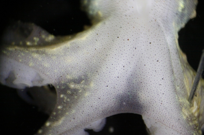
Fig. 2. Posterior side of rubescens during dissection of the mantle, prior to chromatophore activity.

When the radial muscles surrounding chromatophores are relaxed, the elasticity of the tissue retracts the sacculus (Messenger, 2001).  The deceased specimen appeared mostly white to the naked eye (see Fig. 2). It was unexpected to find that individual chromatophores, when observing the epidermis at this magnification, were expanding—the radial muscles around them were contracting. As the solution gradually increased in temperature, this activity increased in frequency and magnitude. Eventually, the posterior side of the specimen was ablaze with changing colors visible to the naked eye.

Video 1. 19:26, solution temperature 12.5°C                               

Video 2. Magnified. 19:32, solution temperature 12.5°C.

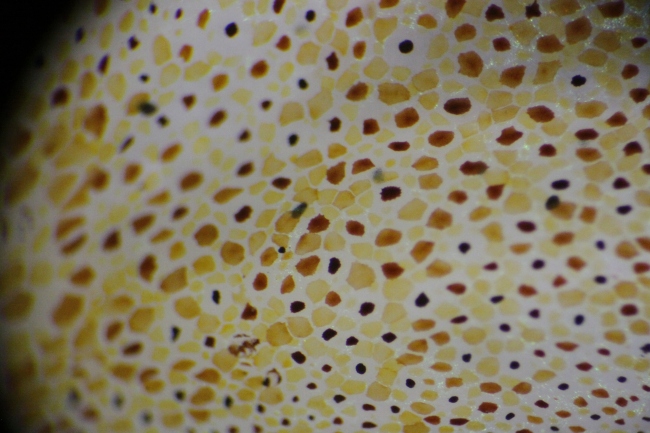
Pigment within the sacculus generally falls toward higher wavelengths of the visible spectrum. Yellow, orange, red, and brown characterized the range found on the O. rubescens (see Fig. 3). These pigments are typically derivatives of tryptophan (Messenger 2001). The activity was visible in waves, and seemed to involve one chromatophore color at a time. Considerable overlap between individual chromatophores was observed when contracted, which, when seen from a distance, could allow further ranges of color. This, along with light reflecting iridophores and the ability of the octopus to contort its epidermis to varying textures, probably aids the octopus in camouflage.

Image

Fig. 3.   Magnified epidermis of O. rubescens as chromatophore activity escalated.

The specimen was cooled via refrigeration and addition of seawater to the solution. This was done to minimize time between observations so as to minimize natural degeneration as a factor for reduced activity. At a solution temperature of 7.5 °C, 36 minutes after start of cooling, the specimen was again observed. The chromatophore activity had perceptibly reduced. This observation supports that this chromatophore activity increases with higher temperatures.

 Video 3. 20:13, solution temperature 7.5°C.

This activity was known by physiologists as “Wandernwolken” (“wandering clouds”) in the nineteenth century. Messenger explains this phenomenon as correlated with the death of the innervating cells while the surrounding muscles endure, and waves of color as was observed here could be induced via anaesthetization with ethanol. These waves tend to propagate through “classes” of chromatophores, such as similar color or age. This activity lends support to a form of network link between chromatophores. (Messenger, 2001).

Dominic Sivitilli

University of Washington

References:

Cloney, R. A. & Florey, E. (1968). Ultrastructure of cephalopod chromatophore organs. Zeitschrift für Zellforschung 89, 250-280.

Messenger, J. B. (2001), Cephalopod chromatophores: neurobiology and natural history. Biological Reviews, 76: 473–528. doi: 10.1017/S1464793101005772

Beauty may be skin deep, but peculiarity isn’t: a close look at Armina californica (Mollusca, Gastropoda, Arminidae)

Meet Armina californica: 

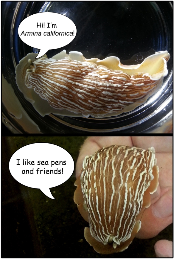
Figure 1: Pleased to meet you!

Figure 1: Pleased to meet you!

If the nudibranch world was a high school, the arminids would be the misfits. With their distinct lack of frills and gills and unique lifestyle, they don’t blend in with any other clique—the fancy aeolids, the sweet dorids (don’t even get me started on those sea-huggin’ dendronotids). However, the arminids don’t seem to mind the exclusion. Measuring up to around 10 cm in length, these sand-cruisin’ sea slugs can easily chomp away on a sea pen alone; but they are often found clustering together sharing a yummy Ptilosarcus meal.

It’s not only their peculiar appearance that sets them apart; their eccentricity runs much deeper than mere physical characteristics. The bauplan of A. californica seems exceedingly simple and inconspicuous, but the internal features make biologists scratch their heads.

External Anatomy

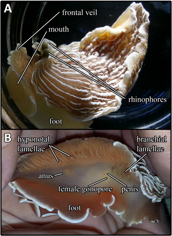
The body of A. californica is typically grayish-pink in color with white peripheral margins and meridional ridges. The striated bulbous rhinophores protrude tightly from a small notch at the anterior dorsum. Unlike the aeolids and dendronotids which have respiratory structures within their cerata and the dorids which have distinctive branchial plumes, the arminids do not possess obvious gills. Instead, the strategy involves sets of lamellae that act as respiratory structures (branchial and hyponotal lamellae) located on the underside of the dorsal flaps. The right side of the foot contains both the anal opening and a gonopore complex that is host to both the female opening and the penis. On the ventral side, the foot extends laterally beyond the edges of the dorsum and anteriorly into a veil-like structure covering the mouth. This gives the animal an overall two-tiered structure.

Figure 2: External anatomy. A) left is anterior; B) right side of the animal (right is anterior).

Figure 2: External anatomy. A) left is anterior; B) right side of the animal (right is anterior).

Internal Anatomy

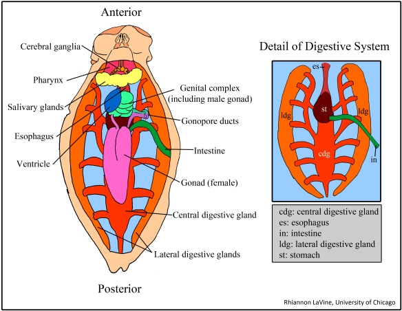
Figure 3: Dorsal dissection with inset of digestive system detail.

Figure 3: Dorsal dissection with inset of digestive system detail.

The dissection of this animal is performed as a simple longitudinal incision of the dorsal surface. The most striking aspect of the internal structures may be the fact that many organs, including the cerebral ganglia, are colored bright orange due to the abundance of carotenoids from the armind’s diet of Ptilosarcus. At the most anterior end, the bright orange 4-ganglia brain sits atop the buccal mass (which contains the pharynx, radular sac, and all related mouthparts). Moving posteriorly, the salivary gland mass conceals the esophagus and its connection to the stomach. A bundle of organs that lies behind the salivary gland includes a clear ventricle and the genital complex—a set of glands and structures that aid in reproduction in addition to the male gonad. Connected to the genital complex is a large, orange gonad which houses the female reproductive components. Both the female gonad and the genital complex are connected via a paired gonoduct to the exterior gonopore complex.

The digestive system includes the anteriormost esophagus which leads directly into the stomach. The stomach extends uninterrupted into the central digestive gland forming one large continuous structure from which emerges the intestine. The branches of the central digestive gland connect back to the stomach, and to a pair of large, orange, lateral digestive glands, the function of which is as of yet unclear and makes this organism quite peculiar. It has been suggested that these glands play a role in the processing of the A. californicus’s odd diet of sea pens, a prey item that can fight back with stinging cells known as nematocytes. Much to the sea pen’s dismay, the arminid seems immune to its venom. The placement of the lateral digestive glands on the body wall may also hint at a further function: are these organs somehow retaining the nematocysts? Perhaps this foreshadows a reuse-recycle defense mechanism similar to that of other gastropods that feed upon cnidarians. The idea is definitely some some food for thought when considering these eccentric bumblers of the sea.

Rhi LaVine
University of Chicago

Native epifaunal communities on invasive Crassostrea gigas (Mollusca, Bivalvia) in Argyle Lagoon

The Pacific Oyster, Crassostrea gigas (Fig. 1), was introduced from Japan to western Washington in the 1920s to replace the dwindling populations of the much smaller, slow-growing native oyster, Ostrea lurida. C. gigas is more robust and economically viable and outcompetes what little population of O. lurida remains in western Washington (White 2009).

big w-scale

Figure 1: Crassostrea gigas individual collected from Argyle Lagoon, San Juan Island, Wa. This non-native species can grow to 45 cm in length, whereas the native Ostrea lurida only grows to approximately 9 cm.

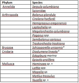
Crassostrea gigas is abundant in the northwest portion of Argyle Lagoon Biological Preserve on San Juan Island, Washington. The oysters are cemented to the small cobbles that make up the bed of the lagoon and are therefore effectively “free living.” The exterior of C. gigas shells are highly fluted with large, irregular furrows and the inner surface is smooth. The unique texture of the valves provides a heterogeneous microhabitat suitable for many of the native organisms that populate the lagoon. During low tide on July 10th, 2013, I collected from Argyle Lagoon ten C. gigas individuals (7 alive, 3 dead) ranging in size from 8.5 to 21 cm in length. I brought them back to the lab where I used Kozloff’s Keys to the Marine Invertebrates and Lamb and Hanby’s Marine Life of the Pacific Northwest to identify all macrofauna living on and within the non-native oyster shells. Diverse communities of organisms representing five metazoan phyla were found on the surface of the ten C. gigas individuals (Table 1).
   

Table

Table 1: Species found on C. gigas shells in Aryle Lagoon.
*Non-native species
+ Only egg masses found (no adults)

Limpets (Lottia spp.) were present on every oyster examined and occurred in densities of up to 26 Lottia per C. gigas individual. Mossy chitons (Moplia spp.), acorn barnacles (Balanus glandula), and burrowing spionid worms were also found on all ten oysters collected from the lagoon (Fig. 2).

anemone

Figure 2: Members of three different phyla (Mollusca, Annelida, and Cnidaria) occupying the same C. gigas shell.

The high abundance of grazers Mopalia and Lottia on the surface of C. gigas is likely due to the presence of algae such as Enteromorpha, Ulva, chain diatoms, and microalgae on the same shells (Fig. 3). Sessile organisms Balanus glandula, Serpula columbiana, and Mytilus trossulus use the C. gigas shells as a hard substrate for settlement while gastropods Haminoea and Nucella use them as sites for laying egg masses. The empty shells of dead Pacific Oysters provide a potentially safe hiding space for the Pagarus spp. and Hemigrapsus oregonensis found within and even more surface area for grazers (Fig. 4).

algae

Figure 3: Surface of C. gigas valve covered with a layer of algae.

dead

Figure 4: Empty C. gigas shell. Small hermit crabs, shore crabs, tanaid shrimp and amphipods were found between the two valves.

Although C. gigas provides substrate for native fauna, contributes to local plankton populations, and is preyed upon by native gastropods and echinoderms, it has been a problematic species for Puget Sound ecosystems because it competitively excludes the native Olympia Oyster from much of its historical range. When C. gigas was introduced to Western Washington for aquaculture, many other non-native Asian species such as Sargassum, Schizoporella unicronis and Ocinebrinus inornata were introduced with it and are now prevalent and in some cases problematic in our marine habitats (White 2009; Lamb and Hanby 2005; Harbo 2011)(Fig. 5).

limpet and bryozoan (1)

Figure 5: C. gigas with encrusting Schizoporella unicornis. S. unicornis was introduced to Western Washington with Pacific Oyster seed (Harbo 2011; Lamb and Hanby 2005).

Sources:
Harbo, R.M. 2011.Whelks to Whales: Coastal Marine Life of the Pacific Northwest. Harbour Publishing Co., Madeira Park, BC, Canada.

Kozloff, E.N. 1974. Keys to the marine invertebrates of Puget Sound, the San Juan Archipelago, and adjacent regions. University of Washington Press, Seattle, Washington.

Lamb, A. & B.P. Hanby. 2005. Marine Life of the Pacific Northwest. Harbour Publishing, Madeira Park, BC, Canada.

White, J., J.L. Ruesink & A.C. Trimble. 2009. The Nearly Forgotten Oyster: Ostrea lurida Carpenter 1864 (Olympia Oyster) History and Management in Wasington State. Journal of Shellfish Research 28(1): 43-49.

Bailey Craig- University of Washington (B.S. 06/2011)