The Decorations of Oregonia gracilis (Decapoda, Brachyura, Majidae)

If you pull up any tire from the edge of the docks at Friday Harbor Laboratories, you’ll find a multitude of organisms — algae, bryozoans, hydroids, tube worms, and tunicates growing on the surface, and many more mobile organisms such as crabs, shrimp, urchins, copepods, and a variety of worms living in the habitat thus created.
DGP_3651

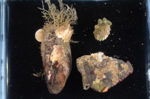
Figure 1 — Mussel (left), limpet (upper right), and rock (lower right), overgrown with a variety of organisms

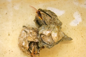
On the benthos, a similar situation exists — any available substrate, living or not, is swiftly colonized by a variety of organisms. The rock in figure 1, though relatively small, is home to Terebratalia transversa, a brachiopod; red algae; at least four species of encrusting bryozoans; small calcareous tubes which could belong to gastropods or serpulid polychaetes; and two larger worm tubes, one containing its worm (Sabellaria), one containing a hermit crab (Discorsopagurus). Living substrates are likewise colonized; the mussel in figure 1, which was dredged from the same location as the rock, is overgrown by Balanus, the acorn barnacle; T. transversa; and at least two types of thecate hydroids. Finally, the limpet (next to the mussel in figure 1) carries on its back the calcareous bryozoan Heteropora, spionid polychaetes, Balanus, red algae, and an encrusting bryozoan. This sort of overgrowth of some organisms by others is an inevitable consequence of the scarcity of space in the marine benthos. Some, such as cheilostome bryozoans with their grabbing avicularia, combat the encrustation. Some, like the mussels and the limpet, ignore it. Finally, some organisms take advantage of it.

The decorator crab (Oregonia gracilis) is named for the patchwork of organisms which it purposefully attaches onto hooked setae on its carapace. Once emplaced, the organisms establish and grow there. This camouflaging cloak of colonists serves to confuse predators, and the makeup of the decorations is known to vary from one place to another, since these crabs choose their decorations from locally-available materials (Wickstein 1992). Decorator crabs are commonly seen on the support posts of the dock here at Friday Harbor, below the water line, so I went down to the dock with a net and a bucket to catch a few and see what they had attached to their carapaces.

I caught three O. gracilis individuals. My first impression of their decorations was that in contrast to the rock, mussel, and limpet I examined earlier, the crabs’ encrusting biota was much more uniform. While the rock had patches of bryozoans and clumps of worm tubes, and the mussel’s hydroids were concentrated at one end, the crabs had organisms evenly distributed over their carapaces. Furthermore, the community composition of different parts of a crab’s carapace was similar; crabs with bryozoans on their backs also had the same type of bryozoans on their legs (Figs. 2, 4), and a crab with spots of sponge on its back had spots of the same sponge on its legs (Fig. 3). In contrast, the rock and mussel communities were much patchier.

So, what lives on the carapaces of decorator crabs at Friday Harbor? Let’s take a look.

Fig2

Figure 2 — Dorsal view of O. gracilis with Dendrobenia lichenoides, red algae, yellow sponge, Corella willmeriana (transparent tunicate), four hydroids, and diatoms

The first crab I caught (Fig. 2) had attached a large frill of the bryozoan Dendrobaenia lichenoides to its rostrum. Smaller flakes of D. lichenoides were attached to its carapace and legs. Among the bryozoans grew at least four species of colonial hydroids. Diatom chains were stuck among the hydroids in many places; since those were not attached directly to the carapace, I am not sure whether or not the crab did that on purpose. Finally, the crab had finished off its decorations with a few bits of red algae, some unidentified yellow sponge, and a transparent tunicate which it carried along on one leg.
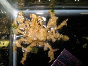
Fig3

Figure 3 — Dorsal view of O. gracilis with Dendrobenia lichenoides, green algae, red algae, and unidentified hydroids

The second crab (Fig. 3), smaller than the first, was thickly adorned with D. lichenoides and a number of hydroids. It added some heterogeneity into its camouflage with some flecks of red and green algae.
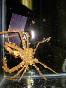
Fig4

Figure 4 — Dorsal view of O. gracilis with Dendrobenia lichenoides, yellow sponge, red algae, and green algae

The third crab (Fig. 4), like the first two, had also placed D. lichenoides all over its body. Unlike the first two, however, it had also polka-dotted itself with yellow sponges. It also had a few flecks of red and green algae, but no hydroids that I could find.

While the species composition from one crab’s carapace to another is not identical, a number of similarities emerge. Overall, the crabs appear to have attached leafy or dendritic, neutral-colored, colonial animals over most of their carapaces, and then added smaller patches of brighter-colored materials. The species on their carapaces are all common on local non-living substrates; for instance, it is easy to find Dendrobaenia on the dock tires. Finally, just like the bryozoans, algae, and hydroids on the dock tires and the dredged rocks and mussel, the habitat built by the decorator crabs on their carapaces provides a home for a number of other species; I found a number of amphipods, nematodes, and polychaetes living amongst their bryozoans and hydroids. Thus, while decorator crabs purposefully cultivate sessile organisms for their camouflage, they incidentally attract more mobile organisms as well and thus carry full-fledged communities on their backs.

Nadia Pierrehumbert
University of Chicago

REFERENCES

Wicksten, Mary K. 1993. A review and a model of decorating behavior in spider crabs (Decapoda, Brachyura, Majidae). Crustaceana 64(3).

Sampling the Diversity of Alpheid Shrimp Microhabitats: Examples from the Pacific Northwest and the Indo-West Pacific

Alpheidae is a family of caridean shrimp that contains the snapping shrimp genera Alpheus and Synalpheus, in addition to a few other non-snapping genera. Although the most diverse genus in this family (Alpheus) typically inhabits nearly all tropical and sub-tropical shallow water marine habitats, alpheids in general can be found in many diverse places, from mud flats to coral reefs to seagrass beds, including in temperate waters, with some individuals even inhabiting deep-water Caribbean locales.

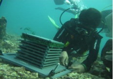
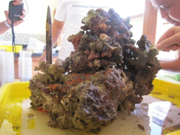
I experienced a small portion of this habitat diversity during two field seasons studying Alpheus genetic and cryptic diversity in Bali and Sumatra, Indonesia. Most of our samples were collected from the interstices of dead Pocillopora spp. coral heads (Fig. 1) that were excavated from the reefs, and all others were taken from the surfaces of our Artificial Reef Monitoring Structures (ARMS) (Fig. 2) that were placed on the reefs the year before. During this time, we collected shrimp from 47 unique evolutionarily significant units (ESUs) from many different types of microhabitats on both the ARMS and the coral heads.

Figure 1: Dead Pocillopora coral head taken from a reef off the shore of northern Bali, Indonesia. All Alpheus samples taken from the head were found in the interstices of the coral (from both living and dead colonies), inside of algal tubes hanging from the head, or within caverns excavated inside of the skeleton itself.

Figure 1: Dead Pocillopora coral head taken from a reef off the shore of northern Bali, Indonesia. All Alpheus samples taken from the head were found in the interstices of the coral (from both living and dead colonies), inside of algal tubes hanging from the head, or within caverns excavated inside of the skeleton itself.

Figure 2: Artificial Reef Monitoring Structure (ARMS) being deployed on a reef flat in Bali, Indonesia. All Alpheus samples collected from the ARMS were pulled from the plastic green scrubby shown here as the uppermost tier of the structure.

Figure 2: Artificial Reef Monitoring Structure (ARMS) being deployed on a reef flat in Bali, Indonesia. All Alpheus samples collected from the ARMS were pulled from the plastic green scrubby shown here as the uppermost tier of the structure.

One of the many sampled microhabitats was within the crevices of living Pocillopora spp. corals; A. lottini is a mutualistic symbiont of corals of this genus, trading its defensive capabilities in return for shelter (Fig. 3). It has also adopted cryptic coloration patterns for additional protection from predators.

Figure 3: A. lottini individual collected from a section of living Pocillopora coral growing over a dead coral head. It is a mutualistic symbiont of the living corals and its coloration is cryptic among the colony's interstices.

Figure 3: A. lottini individual collected from a section of living Pocillopora coral growing over a dead coral head. It is a mutualistic symbiont of the living corals and its coloration is cryptic among the colony’s interstices.

Another microhabitat we sampled was inhabited by A. frontalis: shrimp of this species formed tubes from algal mats that grew over the dead corals (Fig. 4).

Figure 4: A. frontalis individual collected from within algal tubes it made from algal mats overgrowing the dead coral skeleton.

Figure 4: A. frontalis individual collected from within algal tubes it made from algal mats overgrowing the dead coral skeleton.

A. obesomanus, which is recognizable by the distinctive hammer-like dactylus on its major claw, was found within tunnels it had excavated inside the coral’s limestone skeleton using this modified dactylus (Fig. 5).

Figure 5: A. obesomanus ovigerous female collected from a cavity she excavated inside of the dead coral skeleton using the hammer-like dactylus of her major cheliped. She is shown with a bopyrid parasite inside of her carapace.

Figure 5: A. obesomanus ovigerous female collected from a cavity she excavated inside of the dead coral skeleton using the hammer-like dactylus of her major cheliped. She is shown with a bopyrid parasite inside of her carapace.

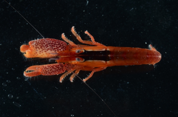
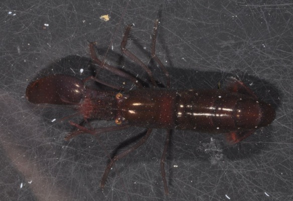
We also observed shrimps in the genus Synalpheus living with sea lilies – individuals we collected from crinoids of different colors were found to be of the same species complex (S. stimpsonii), but were cryptically colored to match their hosts (Fig. 6).

Figure 6a: Ovigerous female of the S. stimpsonii complex collected from the oral surface of the central disk of a yellow crinoid.

Figure 6a: Ovigerous female of the S. stimpsonii complex collected from the oral surface of the central disk of a yellow crinoid.

Figure 6b: S. stimpsonii male collected alongside (6a) on a yellow crinoid.

Figure 6b: S. stimpsonii male collected alongside (6a) on a yellow crinoid.

Figure 6c: Ovigerous female of the S. stimpsonii complex collected from a black crinoid on the reef flat.

Figure 6c: Ovigerous female of the S. stimpsonii complex collected from a black crinoid on the reef flat.

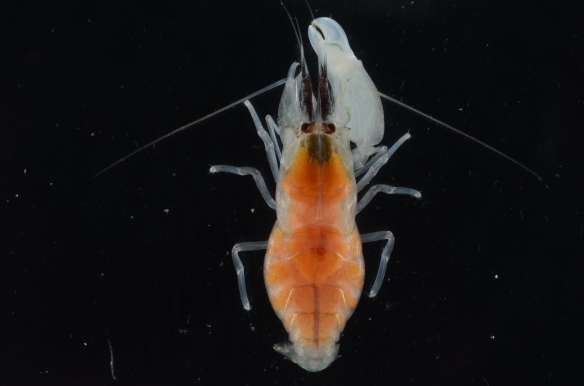
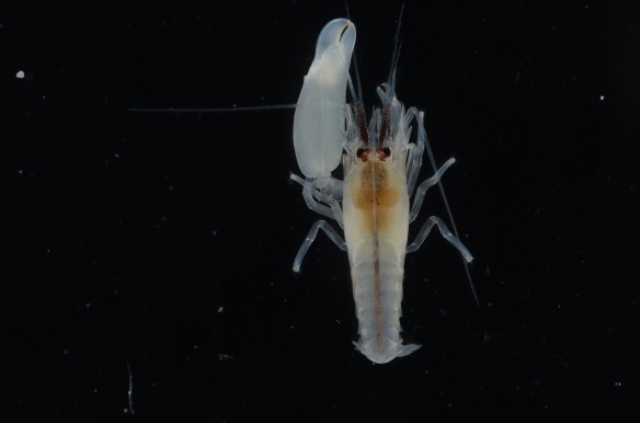
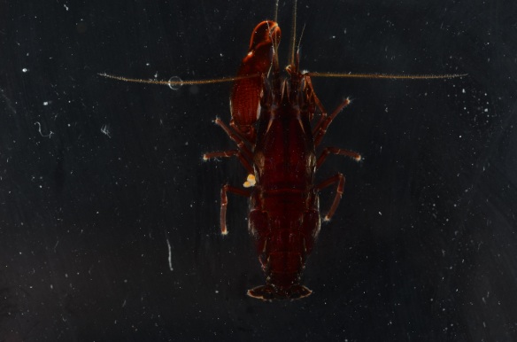
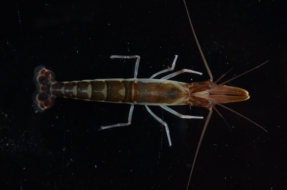
During my time on San Juan Island, I was also fortunate enough to collect a single alpheid representative from the mud flats of Argyle Bay during low tide on July 10, 2013. This individual, of the species Betaeus harrimani, was a commensal of the mud shrimp, Upogebia spp., and was collected from the caverns excavated by the much larger shrimp. The individual I encountered was an ovigerous female that was 4.9cm long (Fig. 7).

Figure 7: Ovigerous B. harrimani female collected from the cavern system dug by Upogebia in the mud flats of Argyle Bay on San Juan Island.

Figure 7: Ovigerous B. harrimani female collected from the cavern system dug by Upogebia in the mud flats of Argyle Bay on San Juan Island.

Betaeus is a minor genus of Alpheidae, containing around 28 species, and in the Pacific Northwest, there are only representatives from two of these species. What surprised me most was that this genus does not possess asymmetrical chelipeds and cannot snap. During my time working with the Betaeus female, I found that instead of snapping to defend herself, like all of the other alpheid shrimp I’ve worked with, she held out her symmetrical pincers in a defensive stance that resembled that employed by many Brachyuran crabs when threatened.

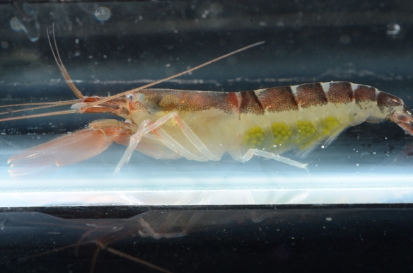
While observing her behavior, I also noticed that she used both her pleopods and her pereopods to locomote. This struck me as odd because I knew she was ovigerous and that caridean shrimp attach their eggs to their pleopods via an adhesive substance. Upon a closer look at her swimming legs, it appeared that the eggs were attached instead to her sternites since the legs and the eggs moved independently while she swam (Fig. 8). In terms of both aerating the eggs and having them remain attached to the body of the mother during development, it would seem as if having the eggs attached to the sternites instead of the swimming legs would be evolutionarily more advantageous.

Figure 8: Left lateral of B. harrimani female. While the pleopods are stationary, the eggs appear to be attached to the legs themselves, but while swimming, the eggs move independently of the pleopods.

Figure 8: Left lateral of B. harrimani female. While the pleopods are stationary, the eggs appear to be attached to the legs themselves, but while swimming, the eggs move independently of the pleopods.

To conclude, in terms of habitat similarities between all of the alpheids I’ve collected, I would hypothesize that shrimp in this family prefer to live interstitially; every shrimp I have collected was found in some sort of nook, be it made of coral, algae, sponge tissue, echinoderm appendages, plastic (in the case of the “scrubby” layer on the ARMS), or gravel. When removed from this habitat and placed into a dish lacking any sort of overhead coverage, each shrimp tended to display thigmotaxis towards the edges of the container. So although the diversity of alpheid habitat preference is great, environmental coverage appears to be the thread that ties them all together.

Victoria Morgan; Cornell University c/o 2013

Color variation in Hemigrapsus oregonensis (Arthropoda, Pancrustacea, Malacostraca, Brachyura)

crab anatomy

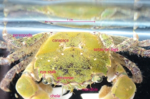
Fig. 1: Anterior-dorsal view of Hemigrapsus oregonensis with labeled external anatomy

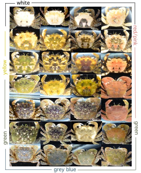
Though Hemigrapsus oregonensis is commonly known as the Green Shore Crab, it does not always seem to be particularly green. This small crab (fig. 1), found along the Pacific coast from Alaska to southern California, can be any number of colors (see fig. 2). Among 67 specimens collected from Argyle Creek on San Juan Island, WA, and analyzed at FHL, the following color patterns emerged: the crabs seemed to have a solid color on their carapace with a distinct pattern of spots or shapes in another color over this. The solid colors in the sampled individuals were white, red, yellow, grey-blue and many shades of green. Often the carapace color would be a blend or mixture of two or three of these colors. The patterns were usually in black or green, often with small white spots. The setose pereopods of the crabs, which distinguishes them from the similar looking Hemigrapsus nudus, were often green with black and white spots, though they could be any color. Chelae were often white.

Image

Fig. 2: Photos of selected crabs’ carapaces. All collected crabs fit into one of these 28 categories. Of 67 specimens, 28 color morphs were found which suggests that there are likely many more variations out there.

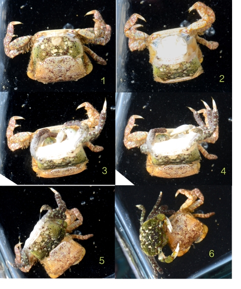
The color variation among this species could be due to genetic variation, sexual dimorphism, environmental factors during or after development, or could change with age or molting stage of the crab. This last hypothesis is supported by evidence that was fortuitously gathered when one crab molted while it was being photographed. The crab was originally beige with maroon and white spots, but after ecdysis the individual emerged with a bright green carapace and the exact same spot pattern as before. This could indicate that color changes with new molts, or that as the crab approaches its molting period, the carapace fades. However, many individuals were lighter than the crab that molted, and did not molt after seven days in the lab (though the environment could have prevented them). Some molted carapaces were found of various colors, many bright red, which seems to contradict the hypothesis that carapace brightness and color are indicative of molting stage.

Image

Fig. 3: Crab molting. This process took about one minute.

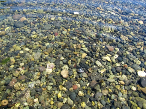
Color also does not seem to depend on age or sex. Individuals were collected from 0.8-2.3 cm, presumably of different ages, with no evidence of a progression or trend in carapace color. There also seemed to be no difference in the colors or patterns between males, females or gravid females. As for habitat, the crabs live among rocks that are just as diversely colored as they are, which likely supports carapace color variation. Crabs did not seem to live under rocks that resembled themselves, and differently colored crabs were often found living under the same rock. Environmental factors likely influence color, but apart from habitat, it is hard to know many details of each crab’s environment, such as their developmental circumstances and diet. Genetics must have some effect on the color, and likely determines the pattern on the carapace because the molting crab kept the same pattern even when its color changed after ecdysis. Most likely, some combination of the above factors must influence carapace color and pattern of Hemigrapsus oregonensis crabs; more research is required to unravel this mystery.

Image

Fig. 4: Habitat of Hemigrapsus oregonensis at Argyle Creek on San Juan Island

Rachel Folz- University of Chicago

Feeding Behavior of Balanus nubilus

Balanus nubilus (Arthropoda, Crustacea, Thecostraca, Cirripedia, Thoracica, Sessilia)

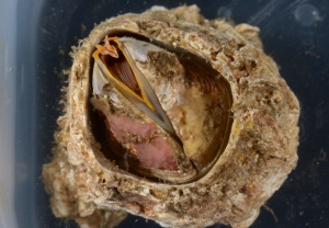
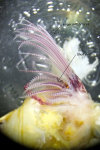
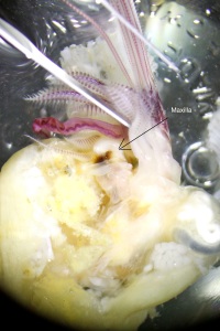
The giant barnacle, Balanus nubilus, is a sessile organism that attaches itself on a chosen substrate and remains there for the duration of the organism’s lifespan (Figure 1). They do not leave their shell, yet open it up for life functions such as: feeding, spawning, and respiration. They are suspension feeding animals that use their feet to extract food from the water column. My observations are based upon organisms with apertures or openings that are around 4cm in length. The giant barnacle has long and short cirripedia showing a clear distinction between the two (Figure 2). Under the short cirri are feeding appendages composed of maxillae and a mandible, which looks very sturdy in comparison to the cirri (Figure 3).

Figure 1. Balanus nubilus with open scutum opercular plates.

Figure 1. Balanus nubilus with open scutum opercular plates.

Figure 2. View of cirripedia, feeding appendages of Balanus nublius.

Figure 2. View of cirripedia, feeding appendages of Balanus nublius.

Figure 3. Dissected Balanus nubilus' feeding appendages, a view into the mouth.

Figure 3. Dissected Balanus nubilus‘ feeding appendages, a view into the mouth.

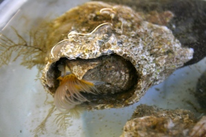
When feeding, the giant barnacle first opens its operculum and then extends and uncurls its cirripedia into the water column (Figure 4). The cirri still retain a slight curve when fully extended into the water column. I found when orienting the water flow from the sea table in the test barnacles direction, verses turing the water flow off, the feeding rate changed. The giant barnacle extends its cirri more frequently when there is less water movement. This appears to increase the water flow around the cirri and helps the barnacle to feed more efficiently. When there is ample water flow, barnacles seem to keep their cirri extended into the water column for longer periods, therefore beating them less frequently. The cirripedia can be swiveled to either side once extended (Figure 5). This action seems to better orient the cirri and face into the current’s flow. Smaller barnacles species feed more rapidly than larger barnacle species. They frequently extend their cirri at a rate of more than once per second whereas larger barnacles beat their cirri at a slower rate.

Figure 4. Ventral view of Balanus nubilus feeding with cirripedia.

Figure 4. Ventral view of Balanus nubilus feeding with cirripedia.

Figure 5. Ventral view of Balanus nubilus orienting cirri in the water column

Figure 5. Ventral view of Balanus nubilus orienting cirri in the water column

Barnacles are likely to stop feeding and narrow the operculum if they sense a predator above them. They seem to most likely sense predators by an interruption of light and shadows being cast over them. An agitated barnacle is timid to readily feed again and may cautiously take many minutes to resume feeding activity.

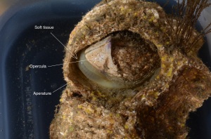
If a barnacle is poked along the soft tissue under the edge of the operculum, it swivels its operculum around the aperture in order to hide the soft and vulnerable tissue from a predator (Figure 6). On large barnacles this sometimes means they expose a vulnerable area of soft tissue on the other side of their opercula. The swiveling movement seems very quick for such a large barnacle.

Figure 6. The soft tissue of Balanus nubilus exposed.

Figure 6. The soft tissue of Balanus nubilus exposed.

Bailey Navratil
University of Washington

The morphology of the Lopholithodes mandtii (Arthropoda, Crustacea, Malacostraca, Decapoda, Anomura)

Members of the Decapoda are crustaceans that have five pairs of walking legs which include the chelipeds, three pairs of maxillipeds that function as mouthparts, two pairs of maxillae, a pair of mandibles which could also be described as jaws for eating, and two pairs of antennae. This general organization varies from species to species.

Crabs are benthic decapods that have a highly reduced abdomen and often a large, wide carapace. “True” crabs are members of the Brachyura.  Members of the Anomura have also evolved crab like features making them a sister group of the Brachyura. A decapod means ten-legged. Brachyura are true crabs because they have five pairs of visible pereiopods, however an Anomura have only four pairs of pereiopods that are visible and a fifth pair that have been reduced to been hiding under the carapace. Lopholithodes mandtii is a large anomuran crab that lives along the pacific northwest cost from Alaska to California and found in the subtidal areas up to 137m (Cowles 2004).  I examined the morphology of one specimen collected subtidally (15 m depth) from Pt. George, Shaw Island.

The most obvious feature of this large crab is that the cuticle of its carapace and legs has large bumps that could make it look or be described as the rough surface of a rock. It seems that this feature of the crab is so that it would be well camouflaged in its rocky habitat. This crab also has a hard, triangular abdomen that is curled under the thorax; it fits into a depression in the thorax but can be pulled away. I examined the appendages of the crab from anterior to posterior.  The first two pairs of appendages are antennae.  The first pair is unbranched (uniramous) and the second pair has two branches (biramous). Behind the antennae is a pair of mandibles, which were hard and rounded, each with a small branch projecting anteriorly.  Posterior to the mandibles are three pairs of appendages that all look similar: they were very thin and leaf-like.  These are called the first and second pairs of maxillae, and the first pair of maxillipeds.  Posterior to those appendages were maxillipeds 2 and 3, which both are biramous bear appendages, modified to function as mouthparts. The first pair of walking legs, which bear  pinching claws, are called chelipeds.  The next three pairs of walking legs have pointed dactyls which are used to grip to hard substrate. At first observation, a fifth pair of pereiopods was not noticeable; however closer observation showed that the reduced fifth pair of pereiopods is hidden under the thorax by the abdomen.

The ecological morphology of the Lopholithodes mandtii is not well studied due to the rareness of its species. However in summary, the morphology of the king crab has been adapted to so that it has the ability to survive and function with ease amongst its rocky deep water environment.

Figure A. Hand drawn diagram of L. mandtii illustration major morphological features of the organism.

Figure 1. Hand drawn diagram of L. mandtii illustrating major morphological features of the organism.

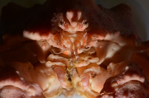
Figure B. Apical view of the organism's mouth parts.

Figure 2. Anterior view of the organism’s mouth parts.

Figure C. Apical view illustrating the asymmetry of the organism.

Figure 3. Anterior view of the organism observing its asymmetry and  its pereopods/dactyl used for hard substrates.

Figure D. Ventral view observing counter shading in the organism L. mandtii.

Figure 4. Ventral view

Figure E. Dorsal view of the L. mandtii observing counter shading.

Figure 5. Dorsal view of the L. mandtii

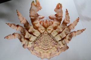
Figure 6. Ventral view of abdomen/thorax, also showing fifth pereiopod reduced

Figure 6. Ventral view of abdomen/thorax, also showing fifth pereiopod reduced

Literature Cited

Cowles, D. Lopholithodes mandtii (Brandt, 1849). 2004. http://www.wallawalla.edu/academics/departments/biology/rosario/inverts/Arthropoda/Crustacea/Malacostraca/Eumalacostraca/Eucarida/Decapoda/Anomura/Family_Lithodidae/Lopholithodes_mandtii.html

Author

Aleisha Setka, Auburn University

Nightlighting at FHL

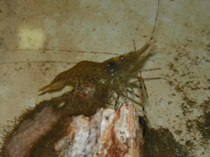
The LED nightlight, just below the surface draws in a couple fast-swimming neirids and a shrimp of the Pandalus genus.

Figure 1: The LED nightlight, just below the surface draws in a couple fast-swimming nereids and a shrimp of the Pandalus genus.

Night Lighting at Friday Harbor Laboratories

By Dan Lombardo

The bulky power supply is connected to the 24” tube of super-bright LEDs, causing students to cry out and cover their eyes, surprised and momentarily stunned by the blinding white flash.

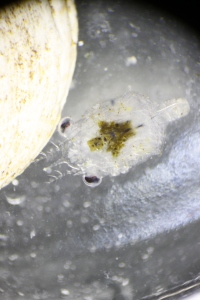
Figure 2: A tiny nauplius larvae that turned out, a few days later, to be a crab of the Cancer genus.

Figure 2: A tiny megalopan larvae that turned out, a few days later, to be a crab of the Cancer genus.

It’s a dark, clear evening, and the nightlight is slowly being lowered off of the floating dock and into the waters of Friday Harbor.  As the afterimages fade, a new world is revealed:  In this world, tiny animals (probably ostracods) race through tight curves beneath the surface like turbo-charged sesame seeds.  The eerie glow of the nightlight is surrounded and smothered in swarms of nauplii (an early larval stage of some crustaceans), while larger and less numerous, the pea-sized final stage of decapod larvae, the megalopa, drift along, their tiny pereopods outstretched in a chibi caricature of adulthood.

Deeper down, just below the nightlight, a churning school of Sandlance, Ammodytes hexapterus, the “potato chip” of the Salish Sea, is beginning to assemble.

As the ebb-tide drains the harbor it brings other planktonic drifters into the submarine halo; Ctenophores (or “comb jellies” if you prefer), of the Bolinopsis and Pleurobrachia genera cruise by, navigating the current with their eight rows of fused cilia in search of prey.  Medusoid hydrozoans, Mitrocoma and Aequoria, like living glass paperweights, pulsate in the moving water column.  As Aqueoria drifts out of the light and toward San Juan Channel, a sudden blue-green flash indicates that its hunt has been successful; its tightly coiled cnidocysts are deployed at amazing speed, impaling and envenomating its planktonic quarry in a horror-movie scene that is thankfully invisible to the naked eye.

100_0182

Figure 3: A coon-stripe shrimp (Pandalus danae) retrieved from the floating dock.

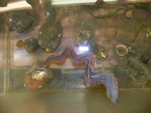
Coon-stripe shrimp, Pandalus danae, crawl, swim, and dart in typical caridoid fashion along the edges of the floating dock.  They are beautifully unaware of our 2-dimensional terrestrial world, and orient themselves in the water column without regard for the illusions of “up” and “down”.  The relative silence is suddenly broken as voices raise to a shout: The creature many of us have been hoping to see is coming into view; writhing and undulating, out of the darkness below, a huge Nereid worm!  The 50cm long giant winds its way through the scene, blindly crashing into the light, the dock, the shrimps, and anything else in its path, in its eagerness to spawn.  This specimen is clearly male, indicated by the milky trail of sperm it leaves behind; the skywriting of a perverted barnstormer, desperately seeking a mate before its terminal disintegration.

Figure 3: The creature everyone wants to see: A massive neirid worm, approaching its spawning phase.

Figure 4: The creature everyone wanted to see: A massive Neanthes brandti!

The best (or easiest to catch) specimens of numerous crustacean larvae, polychaete worms (including our lusty monster Nereid), a few ctenophores, an Aqueoria, and some total unknowns are transferred to our collection jars and buckets and taken to the labs for later evaluation.  It has been a productive evening, and I am looking forward to the chance to practice my diagrammatic drawing during tomorrow’s lab.  While it’s poorly understood why marine animals are drawn to the nightlight, my own attraction is clear:  The nightlight opens the door to an entirely new and fascinating world, a world seldom seen or appreciated by those who aren’t a part of it.  The cryptic nocturnal realm floating just beneath my feet reminds me that there is always more to learn, that the world Is not a simple place, and that the seemingly important complications of my everyday life pale in comparison to the desperate struggles of the quadrillions of tiny creatures more numerous than myself.  It is a scene to be appreciated with awe and humility.

Animals Collected and Identified While Nightlighting in Friday Harbor

Phylum Class Order Family Genus/Species
Annelida Errantia Phyllodocida Nereidae Neanthes Brandti
Arthropoda Malacostraca Decapoda Pandalidae Pandalus danae
  Cancridae Cancer spp.
Cnidaria Hydrozoa Leptomedusae Aequoriedae Aequoria spp.
  Mitrocomidae Mitrocoma cellularia
Ctenophora Tentaculata Cydippida Pleurorachiidae Pleurobrachia bachei
Lobata Bolinopsidae Bolinopsis infundibulum