The Dissection of Abarenicola claparedi (Annelida, Polychaeta)

On a quiet sunny day, my class went to Eagle Cove, San Juan Island to dig Abarenicola claparedi for our first dissection. The best and easiest way to find these worms is looking for their burrow openings and fecal castings on the surface of the sand. Then we used the shovels to dig as much sand as we could and searching them.

lugworm2-2010

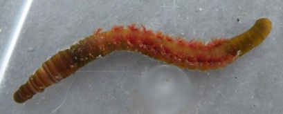
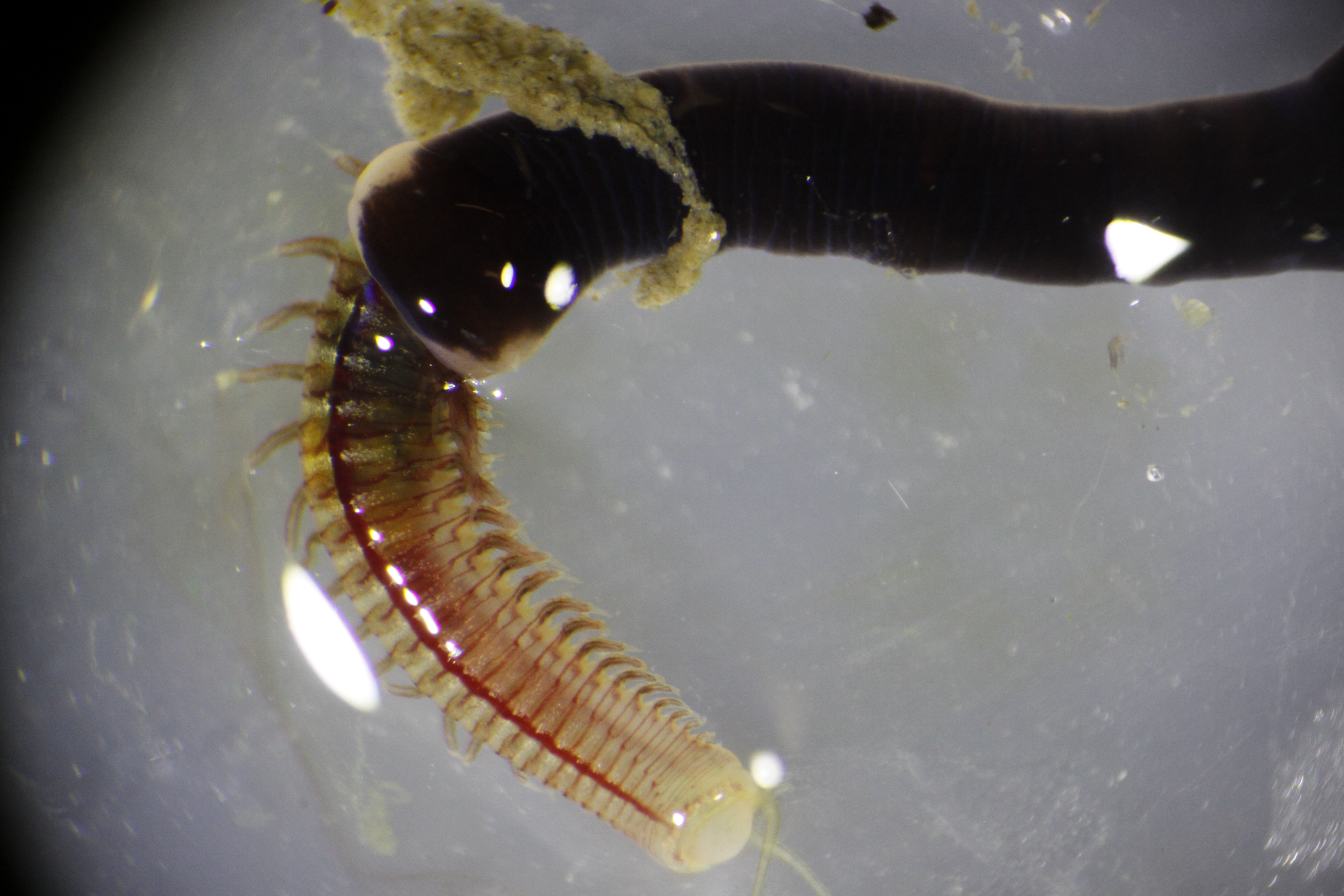
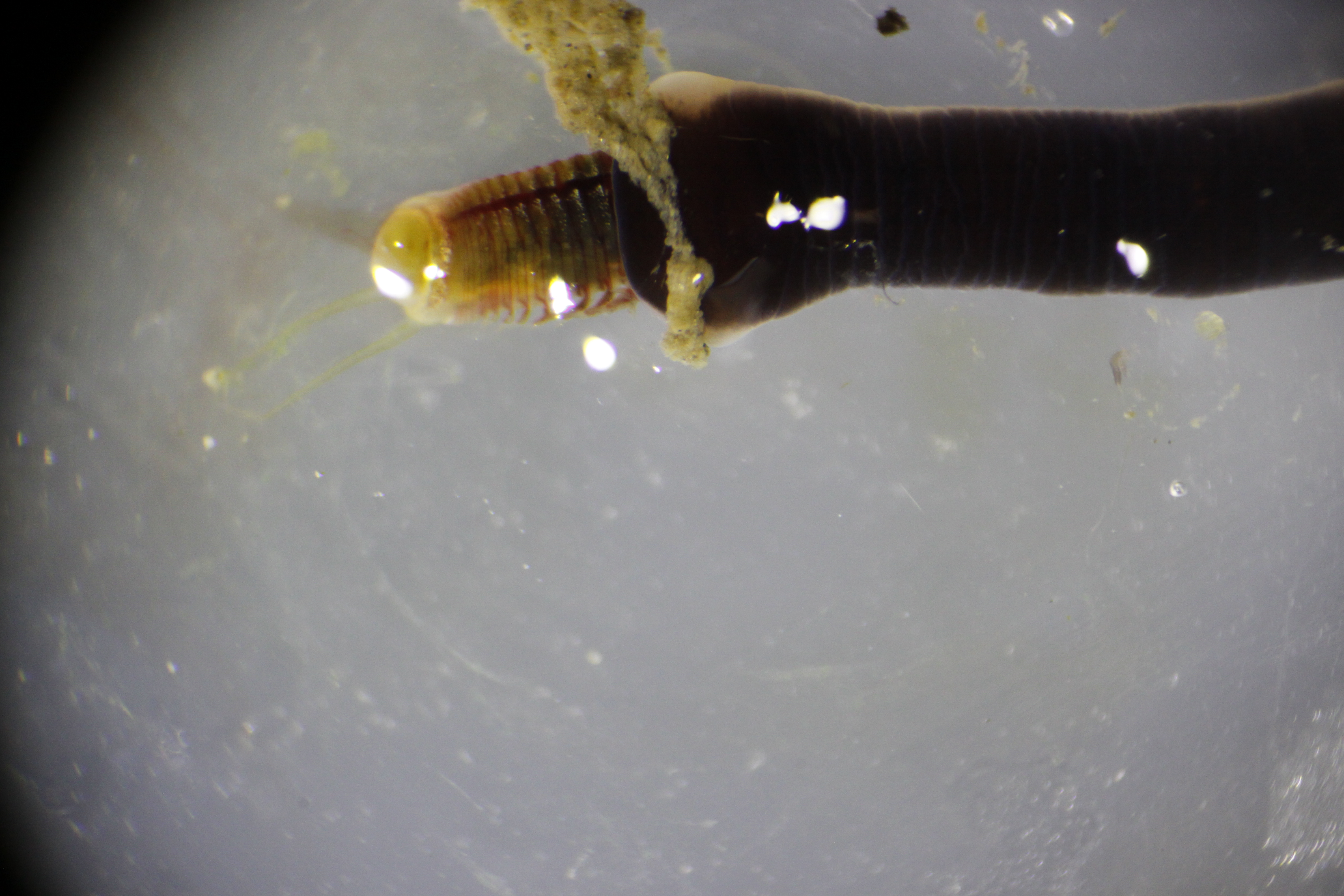
Figure 1. Photo of A. claperedi. Anterior right and posterior left.

1

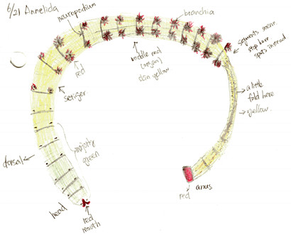
Figure 2. Drawing of live A. claperedi. Dorsal side. Anteiror left, posteiror right.

Abarenicola claparedi belongs to the class Polychaeta, phylum Annelida. Figure 1 and 2 shows the appearance of the A. claperedi. The whole worm is 20cm long, and1cm wide at the head. The first segments, including the head, are green, followed by yellow body segments. The color of the mouth is red and the anus is orange-red. There are red branchia on both sides of the middle region.  A. claperedi lives in borrows in the sand. It swallows the sediment and feeds on the organic material. Its feces can be seen as sandy ropes outside of the opening to its burrow. Meanwhile, the sand will collapse a little because of the swallow.

2

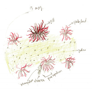
Figure 3. Drawing of dorsal view with branchia. At 15x magnification of dissecting scope. Also shows the complete part of parapodium

Looking closely to the skin, there are orange dots covering the skin. The parapodium consists of a ventral neuropodium with chaetae and a dorsal brachium (gill).  Figure 3 shows the details of the branchia. Lots of branches increase the surface area for gas exchange. The red color is respiratory pigment in capillaries that go through each branch, transferring oxygen while “breathing”. Observed at 15x magnification on the dissecting scope, the skin of the A. claperedi is translucent yellow: you can see the gut but not clearly underneath the skin.

3

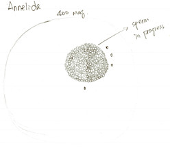
Figure 4. Drawing of a maturing spermatocyte finding in the body liquid of A. claperedi. At 400x maginification of compound scope.

Finishing observations of external features, we started to dissect the A. claperedi. We cut a small hole with fine scissors and sucked some liquid flowing out from the worm. Figure 4 shows what we saw under the compound scope. We didn’t see mature gametes, but think that the cell in figure 4 is a maturing spermatocyte.  The season that A. claperedi spawn sperms is spring and we dissected it in summer.

4

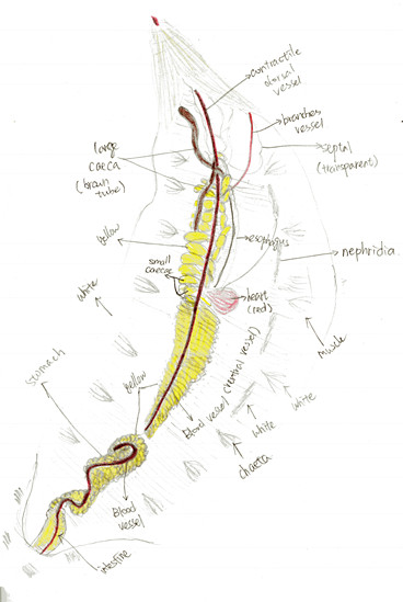
Figure 5. Drawing of the opening body of A. claperedi after dissection. Anterior up, posterior down. Not the compelet worm with part of bottom left. Shows all the organs and muscles. Differenciated by color pencle.

Figure 5 shows the internal structure of A. claperedi. The organs are clear and organized inside its coelom. From the posterior, you can see the intestine, with black sediment in it. It is connected with the stomach. Our worm’s stomach broke in to two parts, maybe in the process of digging it out from the sediment. Anterior to the stomach, you can see the esophageal caeca. There are around 10 to 13 small caeca arranged along the esophagus, posterior to two long, large caeca. Both the stomach and the small caeca are yellow and the large caeca are brown. Between the stomach and the caeca is the heart, in dark red. On the surface of that organ, a relative thick dorsal vessel goes along in the coelom from the anterior to posterior. Furthermore, on the heart side, a line of white nephridia arrange along the skin. You can also see white muscle underneath the skin and also beside the chaetae.

http://www.beachwatchers.wsu.edu/ezidweb/animals/Abarenicolapacifica2.htm

Beauty may be skin deep, but peculiarity isn’t: a close look at Armina californica (Mollusca, Gastropoda, Arminidae)

Meet Armina californica: 

Figure 1: Pleased to meet you!

Figure 1: Pleased to meet you!

If the nudibranch world was a high school, the arminids would be the misfits. With their distinct lack of frills and gills and unique lifestyle, they don’t blend in with any other clique—the fancy aeolids, the sweet dorids (don’t even get me started on those sea-huggin’ dendronotids). However, the arminids don’t seem to mind the exclusion. Measuring up to around 10 cm in length, these sand-cruisin’ sea slugs can easily chomp away on a sea pen alone; but they are often found clustering together sharing a yummy Ptilosarcus meal.

It’s not only their peculiar appearance that sets them apart; their eccentricity runs much deeper than mere physical characteristics. The bauplan of A. californica seems exceedingly simple and inconspicuous, but the internal features make biologists scratch their heads.

External Anatomy

The body of A. californica is typically grayish-pink in color with white peripheral margins and meridional ridges. The striated bulbous rhinophores protrude tightly from a small notch at the anterior dorsum. Unlike the aeolids and dendronotids which have respiratory structures within their cerata and the dorids which have distinctive branchial plumes, the arminids do not possess obvious gills. Instead, the strategy involves sets of lamellae that act as respiratory structures (branchial and hyponotal lamellae) located on the underside of the dorsal flaps. The right side of the foot contains both the anal opening and a gonopore complex that is host to both the female opening and the penis. On the ventral side, the foot extends laterally beyond the edges of the dorsum and anteriorly into a veil-like structure covering the mouth. This gives the animal an overall two-tiered structure.

Figure 2: External anatomy. A) left is anterior; B) right side of the animal (right is anterior).

Figure 2: External anatomy. A) left is anterior; B) right side of the animal (right is anterior).

Internal Anatomy

Figure 3: Dorsal dissection with inset of digestive system detail.

Figure 3: Dorsal dissection with inset of digestive system detail.

The dissection of this animal is performed as a simple longitudinal incision of the dorsal surface. The most striking aspect of the internal structures may be the fact that many organs, including the cerebral ganglia, are colored bright orange due to the abundance of carotenoids from the armind’s diet of Ptilosarcus. At the most anterior end, the bright orange 4-ganglia brain sits atop the buccal mass (which contains the pharynx, radular sac, and all related mouthparts). Moving posteriorly, the salivary gland mass conceals the esophagus and its connection to the stomach. A bundle of organs that lies behind the salivary gland includes a clear ventricle and the genital complex—a set of glands and structures that aid in reproduction in addition to the male gonad. Connected to the genital complex is a large, orange gonad which houses the female reproductive components. Both the female gonad and the genital complex are connected via a paired gonoduct to the exterior gonopore complex.

The digestive system includes the anteriormost esophagus which leads directly into the stomach. The stomach extends uninterrupted into the central digestive gland forming one large continuous structure from which emerges the intestine. The branches of the central digestive gland connect back to the stomach, and to a pair of large, orange, lateral digestive glands, the function of which is as of yet unclear and makes this organism quite peculiar. It has been suggested that these glands play a role in the processing of the A. californicus’s odd diet of sea pens, a prey item that can fight back with stinging cells known as nematocytes. Much to the sea pen’s dismay, the arminid seems immune to its venom. The placement of the lateral digestive glands on the body wall may also hint at a further function: are these organs somehow retaining the nematocysts? Perhaps this foreshadows a reuse-recycle defense mechanism similar to that of other gastropods that feed upon cnidarians. The idea is definitely some some food for thought when considering these eccentric bumblers of the sea.

Rhi LaVine
University of Chicago

Paranemertes peregrina (Nemertea, Hoplonemertea)

Paranemertes peregrina is a species of ribbon worm that inhabits some of San Juan Island’s intertidal zones. I observed these worms in situ and collected them at Eagle Cove and False Bay. The individual pictured in the drawing, one from False Bay, measured approximately 8 cm in length when relaxed and about 11 cm when completely outstretched. In general, most worms that our class collected were about this size, but some ranged up to about 10 cm in length when relaxed. They are dorsoventrally thinned, only measuring 1 to 2 mm in height but about 5 mm in width. Dorsal color is brown/purple, and they have a peach-colored ventral side (see Figure 1). Under a dissecting microscope with a good light source, it is possible to see the coelom-based circulatory system in the form of darker brown lines within the body wall.

Paranemertes1

Figure 1. Dorsal view of a whole P. peregrina specimen, approximately 8 cm long, collected from False Bay.

Movement
These animals are slow movers on land and in water; they use circular and longitudinal muscles arranged down the length of their bodies to inch along sand and rock. The worm that I examined moved by contracting the circular and stretching the longitudinal muscles to elongate its body lengthwise and then doing the opposite to shorten the body, effectively pulling the posterior end anteriorly. At this pulling motion, the anterior end became especially flat, evidently gripping the surface beneath with the added surface area. I saw movement characteristic of only longitudinal muscle action when the individual put its head into the air and moved it side to side, evidently looking to change direction.

Mucus Secretion
When moving, the animal secreted mucus, which could be a mechanism to keep traction with the surface beneath it. I noticed more mucus on the tank walls (smooth, vertical surfaces with little traction) than sand beneath the worm. On the latter, gravity and the rough, moist sand grains aided the animal’s traction. Then, when I picked up the worm it responded by secreting a mucus covering, and I almost lost hold of it. Likely, this would be repeated against any predatory action, enabling the worm to make a slimy escape. In addition to traction and getaway, a mucus covering also helps the worms retain moisture on hot days in the harsh intertidal environment.

Habitat
At False Bay, a muddy intertidal flat, there were hundreds of individuals on the beach at low tide. It was rainy and overcast, so the nemerteans could be on the sand surface without the risk of desiccation. Eagle Cove, the other location, is a rocky intertidal zone. Although there was not nearly the abundance of Paranemertes there (possibly due to the hot weather that day) we found some living on the large rocks in the high intertidal. Although there were not comparable population sizes at the two locations, it is impressive that the species can survive in both muddy and rocky habitats. One factor enabling their habitat diversity is the presence of their prey in both of these habitats.

Feeding
P. peregrina feeds on nereid polychaetes that burrow in the intertidal sand or live in the subtidal. In the lab, we put a P. peregrina individual (9 cm long) in a dish with a nereid worm (8 cm long). After a minute or so of the nereid laying in front of P. peregrina‘s mouth, the ribbon worm everted its proboscis and latched onto its prey at the anterior end (Figure 2). At this point the nereid began to thrash, likely due to Paranemertes piercing it and injecting the neurotoxin, anabaseine (Cowles 2009). The Paranemertes individual reacted to the movement by elongating its proboscis more and more until it wrapped around the nereid, trapping it until the anabaseine took hold and the worm ceased to move. The proboscis extended to a length of a few centimeters during this process, about one third of the length of the worm. Finally, it retracted its proboscis and ingested the nereid through its mouth, anterior end first (Figure 2 and 3).

Paranemertes eating

Figure 2. Dorsal view of P. peregrina in the first stage of feeding with its proboscis everted, attacking and wrapping around a nereid worm. This is the point at which Paranemertes injects anabaseine.

Paranemertes Eating (2)

Figure 3. P. peregrina during the ingestion of the nereid (dorsal view). The red line on the nereid is its large dorsal blood vessel, and the posterior end of the worm is the portion still remaining to be eaten. The individual has expanded itself slightly to ingest this worm, visible by the peach colored ventral side being slightly visible in this dorsal view.

Paranemertes finishing

Figure 3. P. peregrina finishing the ingestion of the nereid (dorsal view). The feces of the nereid is visible at its anus; This Paranemertes individual squeezed the worm so that a large amount of its feces was deposited exteriorly and not consumed.

Maggie Doolin, Hamilton College

Cowles, Dave. Paranemertes peregrina. Walla Walla University, n.d. Web. 11 Jul 2013.

The morphology of the Lopholithodes mandtii (Arthropoda, Crustacea, Malacostraca, Decapoda, Anomura)

Members of the Decapoda are crustaceans that have five pairs of walking legs which include the chelipeds, three pairs of maxillipeds that function as mouthparts, two pairs of maxillae, a pair of mandibles which could also be described as jaws for eating, and two pairs of antennae. This general organization varies from species to species.

Crabs are benthic decapods that have a highly reduced abdomen and often a large, wide carapace. “True” crabs are members of the Brachyura.  Members of the Anomura have also evolved crab like features making them a sister group of the Brachyura. A decapod means ten-legged. Brachyura are true crabs because they have five pairs of visible pereiopods, however an Anomura have only four pairs of pereiopods that are visible and a fifth pair that have been reduced to been hiding under the carapace. Lopholithodes mandtii is a large anomuran crab that lives along the pacific northwest cost from Alaska to California and found in the subtidal areas up to 137m (Cowles 2004).  I examined the morphology of one specimen collected subtidally (15 m depth) from Pt. George, Shaw Island.

The most obvious feature of this large crab is that the cuticle of its carapace and legs has large bumps that could make it look or be described as the rough surface of a rock. It seems that this feature of the crab is so that it would be well camouflaged in its rocky habitat. This crab also has a hard, triangular abdomen that is curled under the thorax; it fits into a depression in the thorax but can be pulled away. I examined the appendages of the crab from anterior to posterior.  The first two pairs of appendages are antennae.  The first pair is unbranched (uniramous) and the second pair has two branches (biramous). Behind the antennae is a pair of mandibles, which were hard and rounded, each with a small branch projecting anteriorly.  Posterior to the mandibles are three pairs of appendages that all look similar: they were very thin and leaf-like.  These are called the first and second pairs of maxillae, and the first pair of maxillipeds.  Posterior to those appendages were maxillipeds 2 and 3, which both are biramous bear appendages, modified to function as mouthparts. The first pair of walking legs, which bear  pinching claws, are called chelipeds.  The next three pairs of walking legs have pointed dactyls which are used to grip to hard substrate. At first observation, a fifth pair of pereiopods was not noticeable; however closer observation showed that the reduced fifth pair of pereiopods is hidden under the thorax by the abdomen.

The ecological morphology of the Lopholithodes mandtii is not well studied due to the rareness of its species. However in summary, the morphology of the king crab has been adapted to so that it has the ability to survive and function with ease amongst its rocky deep water environment.

Figure A. Hand drawn diagram of L. mandtii illustration major morphological features of the organism.

Figure 1. Hand drawn diagram of L. mandtii illustrating major morphological features of the organism.

Figure B. Apical view of the organism's mouth parts.

Figure 2. Anterior view of the organism’s mouth parts.

Figure C. Apical view illustrating the asymmetry of the organism.

Figure 3. Anterior view of the organism observing its asymmetry and  its pereopods/dactyl used for hard substrates.

Figure D. Ventral view observing counter shading in the organism L. mandtii.

Figure 4. Ventral view

Figure E. Dorsal view of the L. mandtii observing counter shading.

Figure 5. Dorsal view of the L. mandtii

Figure 6. Ventral view of abdomen/thorax, also showing fifth pereiopod reduced

Figure 6. Ventral view of abdomen/thorax, also showing fifth pereiopod reduced

Literature Cited

Cowles, D. Lopholithodes mandtii (Brandt, 1849). 2004. http://www.wallawalla.edu/academics/departments/biology/rosario/inverts/Arthropoda/Crustacea/Malacostraca/Eumalacostraca/Eucarida/Decapoda/Anomura/Family_Lithodidae/Lopholithodes_mandtii.html

Author

Aleisha Setka, Auburn University

Native epifaunal communities on invasive Crassostrea gigas (Mollusca, Bivalvia) in Argyle Lagoon

The Pacific Oyster, Crassostrea gigas (Fig. 1), was introduced from Japan to western Washington in the 1920s to replace the dwindling populations of the much smaller, slow-growing native oyster, Ostrea lurida. C. gigas is more robust and economically viable and outcompetes what little population of O. lurida remains in western Washington (White 2009).

big w-scale

Figure 1: Crassostrea gigas individual collected from Argyle Lagoon, San Juan Island, Wa. This non-native species can grow to 45 cm in length, whereas the native Ostrea lurida only grows to approximately 9 cm.

Crassostrea gigas is abundant in the northwest portion of Argyle Lagoon Biological Preserve on San Juan Island, Washington. The oysters are cemented to the small cobbles that make up the bed of the lagoon and are therefore effectively “free living.” The exterior of C. gigas shells are highly fluted with large, irregular furrows and the inner surface is smooth. The unique texture of the valves provides a heterogeneous microhabitat suitable for many of the native organisms that populate the lagoon. During low tide on July 10th, 2013, I collected from Argyle Lagoon ten C. gigas individuals (7 alive, 3 dead) ranging in size from 8.5 to 21 cm in length. I brought them back to the lab where I used Kozloff’s Keys to the Marine Invertebrates and Lamb and Hanby’s Marine Life of the Pacific Northwest to identify all macrofauna living on and within the non-native oyster shells. Diverse communities of organisms representing five metazoan phyla were found on the surface of the ten C. gigas individuals (Table 1).
   

Table

Table 1: Species found on C. gigas shells in Aryle Lagoon.
*Non-native species
+ Only egg masses found (no adults)

Limpets (Lottia spp.) were present on every oyster examined and occurred in densities of up to 26 Lottia per C. gigas individual. Mossy chitons (Moplia spp.), acorn barnacles (Balanus glandula), and burrowing spionid worms were also found on all ten oysters collected from the lagoon (Fig. 2).

anemone

Figure 2: Members of three different phyla (Mollusca, Annelida, and Cnidaria) occupying the same C. gigas shell.

The high abundance of grazers Mopalia and Lottia on the surface of C. gigas is likely due to the presence of algae such as Enteromorpha, Ulva, chain diatoms, and microalgae on the same shells (Fig. 3). Sessile organisms Balanus glandula, Serpula columbiana, and Mytilus trossulus use the C. gigas shells as a hard substrate for settlement while gastropods Haminoea and Nucella use them as sites for laying egg masses. The empty shells of dead Pacific Oysters provide a potentially safe hiding space for the Pagarus spp. and Hemigrapsus oregonensis found within and even more surface area for grazers (Fig. 4).

algae

Figure 3: Surface of C. gigas valve covered with a layer of algae.

dead

Figure 4: Empty C. gigas shell. Small hermit crabs, shore crabs, tanaid shrimp and amphipods were found between the two valves.

Although C. gigas provides substrate for native fauna, contributes to local plankton populations, and is preyed upon by native gastropods and echinoderms, it has been a problematic species for Puget Sound ecosystems because it competitively excludes the native Olympia Oyster from much of its historical range. When C. gigas was introduced to Western Washington for aquaculture, many other non-native Asian species such as Sargassum, Schizoporella unicronis and Ocinebrinus inornata were introduced with it and are now prevalent and in some cases problematic in our marine habitats (White 2009; Lamb and Hanby 2005; Harbo 2011)(Fig. 5).

limpet and bryozoan (1)

Figure 5: C. gigas with encrusting Schizoporella unicornis. S. unicornis was introduced to Western Washington with Pacific Oyster seed (Harbo 2011; Lamb and Hanby 2005).

Sources:
Harbo, R.M. 2011.Whelks to Whales: Coastal Marine Life of the Pacific Northwest. Harbour Publishing Co., Madeira Park, BC, Canada.

Kozloff, E.N. 1974. Keys to the marine invertebrates of Puget Sound, the San Juan Archipelago, and adjacent regions. University of Washington Press, Seattle, Washington.

Lamb, A. & B.P. Hanby. 2005. Marine Life of the Pacific Northwest. Harbour Publishing, Madeira Park, BC, Canada.

White, J., J.L. Ruesink & A.C. Trimble. 2009. The Nearly Forgotten Oyster: Ostrea lurida Carpenter 1864 (Olympia Oyster) History and Management in Wasington State. Journal of Shellfish Research 28(1): 43-49.

Bailey Craig- University of Washington (B.S. 06/2011)

Gorgonocephalus eucnemis (Echinodermata, Ophiuroidea)

Image

DSC_0226b

Figure 1. Aboral view of Gorgonocephalus eucnemis.

DSC_0230b

Figure 2. Oral view of Gorgonocephalus eucnemis.

Commonly referred to as a basket star, Gorgonocephalus eucnemis (seen in figures 1 and 2) is one of ten recognized species within the genus Gorgonocephalus and the only one currently known to occur in the San Juan Islands.  This particular genus has been extremely successful in radiating throughout the world’s oceans, occurring from the Antarctic to the Arctic (figure 4 WoRMS, Stöhr & Hansson 2013).  Gorgonocephalus relies on and is characterized by its five bifurcating arms which unlike most ophiuroids, has a skin covering the ossicles.  It uses its arms to secure itself on substrate in areas with a strong current, where it suspension feeds.  The arms that are acting to feed become curled with a mucus covering at the ends to help capture its prey.  In the picture below, it can be observed how this species will strategically position itself in areas of the highest current, even in a small tank.

DSC_0391

Figure 3. Two G. eucnemis, 3 located on top of the inflowing water, 1 positioned up current of the outgoing water.

Picture1

Figure 4. Gorgonocephalus distribution (WoRMS, Stöhr & Hansson 2013)

During my stay at Friday Harbor Labs, I have already had the pleasure to observe three specimens of G. eucnemis (seen in the pictures below) and interestingly enough, each of the three have had distinct morphological differences from one another.  While many species of Ophiuroidae are recognized to have high levels of within-species morphological variation, it is truly interesting to observe how visually different these specimens look.  I was not able to extend all of the arms properly in the photos without risking damage to the specimens but there is little to no variation in this respect.  The main morphological differences between the three specimens lie within the central disc.  Figure 5 and 7 both show specimens that have an inflated circular central disc.  Figure 6 shows a specimen that is much thinner on the oral-aboral axis and is concave between the 5 main bifurcating arms.  All three specimens also exhibit different color patterns of the central disc but color is also known to commonly vary within species of ophiuroids.

DSC_0226b

  Figure 5. Specimen 1, aboral view

DSC_0375b

Figure 6. Specimen 2, aboral view

DSC_0381bFigure 7. Specimen 3, aboral view

All three specimens were adults and approximately the same size.  I hypothesize that the differing shapes of the central disc actually has something to do with the reproductive stage of the 3 specimens.  Figure 5 shows a specimen that has a swollen disc but I hypothesize has gametes filling in its reproductive bursae slits.  The specimen in figure 6 probably is not reproductively mature or currently producing gametes and the specimen in figure 7 currently has open bursae slits, possessing large masses of eggs.  All three specimens, even with morphological differences, key out to G. eucnemis (Bush, 1920).  When I get back to Auburn, I will perform a genetic analysis using standard COI and 16S markers on arm clips from all three individuals to test the level of genetic differentiation.

Just for fun, I included a photo of Gorgonocephalus chilensis (1 of the other 10 species from this genus) that was collected in January from the Amundsen Sea, off of Antarctica during our cruise on the RVIB Nathaniel B. Palmer.  Notice how similar the physical morphology of the central disc is between this species and G. eucnemis. Morphological differences that can be used to distinguish different species of Gorgonocephalus lies within the spines and skin on the central disc.  To be specific, the shape of the spines and where the skin is present or absent, can be used to key out a species.  Subtle morphological differences emphasizes the need to combine standard systematicswith genetics to make sure we are being accurate in our assumptions about current biodiversity.

DSC_0012b

Figure 8. Gorgonocephalus chilensis aboral viewDSC_0013

Figure 9. Gorgonocephalus chilensis oral view.

Bush, Mildred. “Key to the echinoderms of Friday Harbor, Washington.”Publications-Puget Sound Biological Station 2 (1920): 17.

Stöhr, S.; Hansson, H. (2013). Gorgonocephalus Leach, 1815. In: Stöhr, S., O’Hara, T. (Eds) (2013). World Ophiuroidea database. Accessed through: World Register of Marine Species at http://www.marinespecies.org/aphia.php?p=taxdeta.

Matthew P. Galaska

Auburn University

Nightlighting at FHL

The LED nightlight, just below the surface draws in a couple fast-swimming neirids and a shrimp of the Pandalus genus.

Figure 1: The LED nightlight, just below the surface draws in a couple fast-swimming nereids and a shrimp of the Pandalus genus.

Night Lighting at Friday Harbor Laboratories

By Dan Lombardo

The bulky power supply is connected to the 24” tube of super-bright LEDs, causing students to cry out and cover their eyes, surprised and momentarily stunned by the blinding white flash.

Figure 2: A tiny nauplius larvae that turned out, a few days later, to be a crab of the Cancer genus.

Figure 2: A tiny megalopan larvae that turned out, a few days later, to be a crab of the Cancer genus.

It’s a dark, clear evening, and the nightlight is slowly being lowered off of the floating dock and into the waters of Friday Harbor.  As the afterimages fade, a new world is revealed:  In this world, tiny animals (probably ostracods) race through tight curves beneath the surface like turbo-charged sesame seeds.  The eerie glow of the nightlight is surrounded and smothered in swarms of nauplii (an early larval stage of some crustaceans), while larger and less numerous, the pea-sized final stage of decapod larvae, the megalopa, drift along, their tiny pereopods outstretched in a chibi caricature of adulthood.

Deeper down, just below the nightlight, a churning school of Sandlance, Ammodytes hexapterus, the “potato chip” of the Salish Sea, is beginning to assemble.

As the ebb-tide drains the harbor it brings other planktonic drifters into the submarine halo; Ctenophores (or “comb jellies” if you prefer), of the Bolinopsis and Pleurobrachia genera cruise by, navigating the current with their eight rows of fused cilia in search of prey.  Medusoid hydrozoans, Mitrocoma and Aequoria, like living glass paperweights, pulsate in the moving water column.  As Aqueoria drifts out of the light and toward San Juan Channel, a sudden blue-green flash indicates that its hunt has been successful; its tightly coiled cnidocysts are deployed at amazing speed, impaling and envenomating its planktonic quarry in a horror-movie scene that is thankfully invisible to the naked eye.

100_0182

Figure 3: A coon-stripe shrimp (Pandalus danae) retrieved from the floating dock.

Coon-stripe shrimp, Pandalus danae, crawl, swim, and dart in typical caridoid fashion along the edges of the floating dock.  They are beautifully unaware of our 2-dimensional terrestrial world, and orient themselves in the water column without regard for the illusions of “up” and “down”.  The relative silence is suddenly broken as voices raise to a shout: The creature many of us have been hoping to see is coming into view; writhing and undulating, out of the darkness below, a huge Nereid worm!  The 50cm long giant winds its way through the scene, blindly crashing into the light, the dock, the shrimps, and anything else in its path, in its eagerness to spawn.  This specimen is clearly male, indicated by the milky trail of sperm it leaves behind; the skywriting of a perverted barnstormer, desperately seeking a mate before its terminal disintegration.

Figure 3: The creature everyone wants to see: A massive neirid worm, approaching its spawning phase.

Figure 4: The creature everyone wanted to see: A massive Neanthes brandti!

The best (or easiest to catch) specimens of numerous crustacean larvae, polychaete worms (including our lusty monster Nereid), a few ctenophores, an Aqueoria, and some total unknowns are transferred to our collection jars and buckets and taken to the labs for later evaluation.  It has been a productive evening, and I am looking forward to the chance to practice my diagrammatic drawing during tomorrow’s lab.  While it’s poorly understood why marine animals are drawn to the nightlight, my own attraction is clear:  The nightlight opens the door to an entirely new and fascinating world, a world seldom seen or appreciated by those who aren’t a part of it.  The cryptic nocturnal realm floating just beneath my feet reminds me that there is always more to learn, that the world Is not a simple place, and that the seemingly important complications of my everyday life pale in comparison to the desperate struggles of the quadrillions of tiny creatures more numerous than myself.  It is a scene to be appreciated with awe and humility.

Animals Collected and Identified While Nightlighting in Friday Harbor

Phylum Class Order Family Genus/Species
Annelida Errantia Phyllodocida Nereidae Neanthes Brandti
Arthropoda Malacostraca Decapoda Pandalidae Pandalus danae
  Cancridae Cancer spp.
Cnidaria Hydrozoa Leptomedusae Aequoriedae Aequoria spp.
  Mitrocomidae Mitrocoma cellularia
Ctenophora Tentaculata Cydippida Pleurorachiidae Pleurobrachia bachei
Lobata Bolinopsidae Bolinopsis infundibulum